Clinical Trial
. 2022 Dec 8;7(23):e163030.
doi: 10.1172/jci.insight.163030.

A randomized, phase II study of sequential belimumab and rituximab in primary Sjögren's syndrome

Affiliations
Clinical Trial

A randomized, phase II study of sequential belimumab and rituximab in primary Sjögren's syndrome

Xavier Mariette et al. JCI Insight. .

Abstract

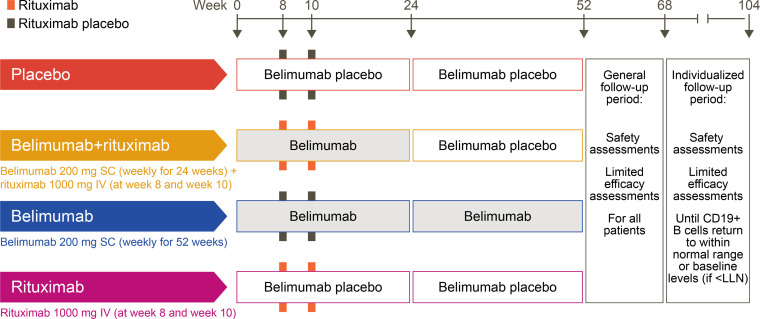
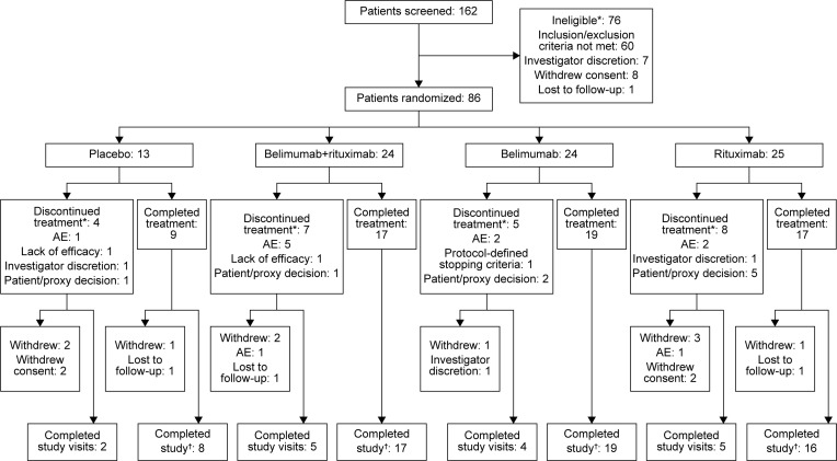
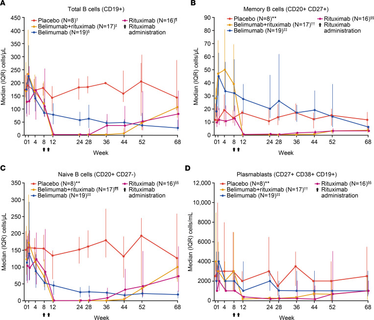
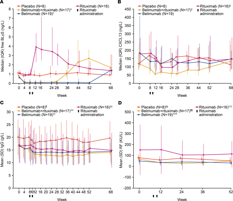
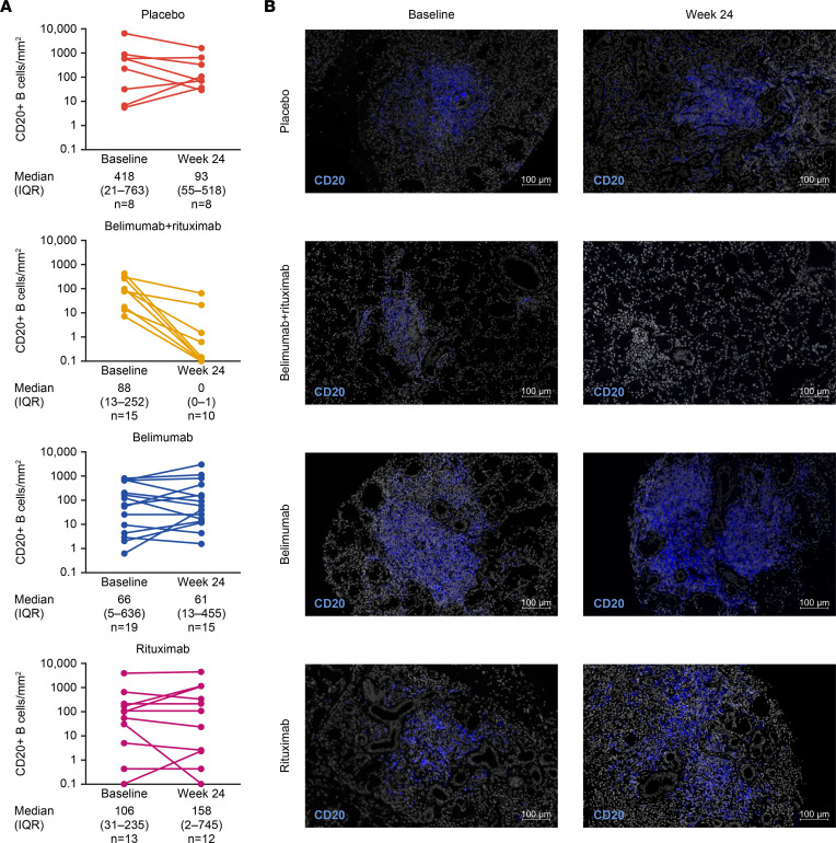
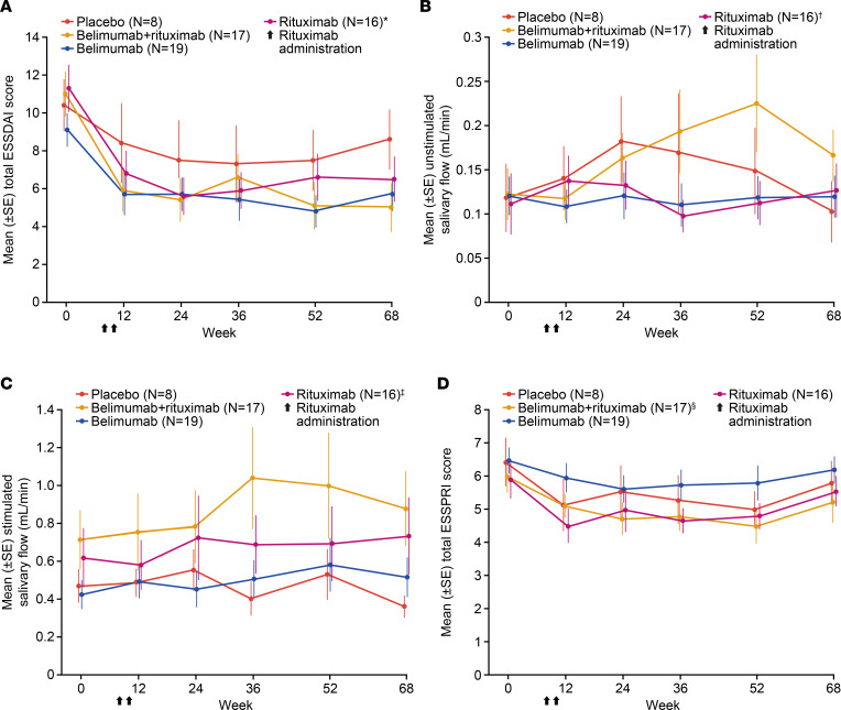
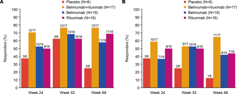
BACKGROUNDPrimary Sjögren's syndrome (pSS) is characterized by B cell hyperactivity and elevated B-lymphocyte stimulator (BLyS). Anti-BLyS treatment (e.g., belimumab) increases peripheral memory B cells; decreases naive, activated, and plasma B cell subsets; and increases stringency on B cell selection during reconstitution. Anti-CD20 therapeutics (e.g., rituximab) bind and deplete CD20-expressing B cells in circulation but are less effective in depleting tissue-resident CD20+ B cells. Combined, these 2 mechanisms may achieve synergistic effects.METHODSThis 68-week, phase II, double-blind study (GSK study 201842) randomized 86 adult patients with active pSS to 1 of 4 arms: placebo, s.c. belimumab, i.v. rituximab, or sequential belimumab + rituximab.RESULTSOverall, 60 patients completed treatment and follow-up until week 68. The incidence of adverse events (AEs) and drug-related AEs was similar across groups. Infections/infestations were the most common AEs, and no serious infections of special interest occurred. Near-complete depletion of minor salivary gland CD20+ B cells and a greater and more sustained depletion of peripheral CD19+ B cells were observed with belimumab + rituximab versus monotherapies. With belimumab + rituximab, reconstitution of peripheral B cells occurred, but it was delayed compared with rituximab. At week 68, mean (± standard error) total EULAR Sjögren's syndrome disease activity index scores decreased from 11.0 (1.17) at baseline to 5.0 (1.27) for belimumab + rituximab and 10.4 (1.36) to 8.6 (1.57) for placebo.CONCLUSIONThe safety profile of belimumab + rituximab in pSS was consistent with the monotherapies. Belimumab + rituximab induced enhanced salivary gland B cell depletion relative to the monotherapies, potentially leading to improved clinical outcomes.TRIAL REGISTRATIONClinicalTrials.gov NCT02631538.FUNDINGFunding was provided by GSK.

Keywords: Autoimmune diseases; Clinical Trials; Drug therapy; Immunology.


Conflict of interest statement

Conflict of interest: XM has received grant/research support from Ose and has been a paid consultant for AstraZeneca, BMS, Galapagos, GSK, Novartis, Pfizer, and Sanofi. FB has received grant/research support from GSK, UCB, Roche, and Actelion; has been a paid consultant for GSK, UCB, Roche, and Actelion; and has been an employee of Kintai Therapeutics and Candel Therapeutics. HB has received grant/research support from BMS and Roche; has been a paid consultant for BMS, Roche, Novartis, MedImmune, UCB, and Servier; and has been a paid speaker for BMS and Novartis. RBH, PM, RP, AVM, NW, and DAR are employees of GSK and hold shares in the company. PPT, KLC, and MH were employees of GSK and held shares in the company at the time of study design and execution. SDV has been a paid consultant for GSK, Novartis, and Roche. KL has participated in advisory boards for GSK and AstraZeneca. RS has been a paid consultant for GSK, Boehringer, Novartis, Janssen, BMS, and AbbVie; has received support to attend meetings from GSK and Amgen; and has been involved with clinical trials for GSK, Servier, USB, and Novartis. JS was an employee of GSK, holds shares in the company, and is currently an employee of AstraZeneca and holds shares in the company. GSK was involved in study design, collection, analysis, and interpretation of data, as well as publication development.

Figures

Figure 1
Figure 1. Study design.
LLN, lower limit of normal.
Figure 2
Figure 2. Patient flow through the study.
*Patients may only have one primary reason;.†Completer population (patients who completed the 52-week treatment and general follow-up period of the study, including the visit at week 68). AE, adverse event.
Figure 3
Figure 3. Median (IQR).
(AD) Total B cells (CD19+), memory B cells (CD20+CD27+), naive B cells (CD20+CD27), and plasmablasts (CD27+CD38+CD19+) over time by flow cytometry (completer population, n = 60). Flow cytometry data were analyzed using the Hodges-Lehmann method to provide a nonparametric 95% CI for the treatment comparisons of interest. For clear presentation of results, data in AC are presented as cells/μL (with different y axes maximum values), and data in D are presented as cells/mL. N = 7 at weeks 4, 12, and 52. N = 16 at weeks 1, 36, 44, 68. §N = 17 at weeks 1, 44, and 68. N = 18 at weeks 4, 12, 28, and 36. N = 16 at week 52. N = 13 at week 24. **N = 7 at weeks 4, 8, and 12. N = 6 at week 52. ††N = 16 at weeks 1, 8, 44, and 68. N = 15 at week 36. ‡‡N = 17 at weeks 1, 44, and 68. N = 18 at weeks 4, 8, 28, and 36. N = 16 at week 12. N = 15 at week 52. §§N = 13 at week 24. N = 14 at week 36. ¶¶N = 16 at weeks 1, 8, 28, 44, and 68. N = 15 at weeks 24 and 36. IQR, interquartile range.
Figure 4
Figure 4. Key serological biomarkers over time.
(AC) Free BLyS, CXCL13, IgG, and RF (patients positive at baseline) (completer population, n = 60). *N = 16 at week 36. N = 18 at week 28. N = 16 at weeks 36 and 44. §N = 15 at week 24. N = 7 at weeks 4 and 48. **N = 16 at week 8. ††N = 17 at week 20. N = 18 at week 36. ‡‡N = 15 at weeks 10, 24, 40, and 48. N = 14 at week 32. §§N = 6 at weeks 0, 36, and 52. N = 7 at week 12. N = 5 at week 24. ¶¶N = 7 at week 0. N = 6 at week 12. N = 5 at weeks 24, 36, and 52. ***N = 14 at week 0. N = 12 at week 12. N = 10 at weeks 24 and 36. N = 9 at week 52. †††N = 11 at week 0. N = 9 at weeks 12, 24, and 36. N = 10 at week 52. BLyS, B-lymphocyte stimulator; CXCL13, chemokine (C-X-C motif) ligand 13; IgG, immunoglobulin G; RF, rheumatoid factor.
Figure 5
Figure 5. Absolute CD20+ B cells in the salivary gland and representative immunofluorescence images.
(A and B) Absolute CD20+ B cells in the salivary gland and representative immunofluorescence (Hoechst CD20) histological images. (completer population, n = 60). Median (IQR): includes all baseline/week 24 completer data. Only data for patients with paired baseline/week 24 biopsies. Minimum values = 0.1. When CD20+ B cells were undetectable, values were input as 0.1 to allow logarithmic display. Changes in absolute CD20+ B cells in the salivary gland were analyzed using the Hodges-Lehmann method to provide a 95% CI for treatment comparisons of interest. For the histological images, original slides were imaged at 20× using a Zeiss Axio Scan Z1 slide scanner and are included in Supplemental Figure 3. IQR, interquartile range. Scale bars: 100 µm.
Figure 6
Figure 6. Clinical efficacy over time as measured by mean (standard error).
(AD) ESSDAI total score, unstimulated salivary flow, stimulated salivary flow, and ESSPRI total score (completer population, n = 60). *N = 15 at week 12. N = 15 at week 36. N = 15 at weeks 36 and 68. §N = 16 at week 52. ESSDAI, EULAR Sjögren’s syndrome disease activity index; ESSPRI, EULAR Sjögren’s Syndrome Patient Reported Index.
Figure 7
Figure 7. Proportion of responders with an ESSDAI reduction.
(A and B) Proportion of responders with an ESSDAI reduction of ≥3 points and ≥5 points versus baseline (completer population, n = 60). ESSDAI responder analyses utilized a generalized estimating equation model. ESSDAI, EULAR Sjögren’s syndrome disease activity index.

References

    1. Del Papa N, Vitali C. Management of primary Sjogren’s syndrome: recent developments and new classification criteria. Ther Adv Musculoskelet Dis. 2018;10(2):39–54. doi: 10.1177/1759720X17746319. - DOI - PMC - PubMed
    1. Omdal R, et al. Pain and fatigue in primary Sjögren’s syndrome. Rheumatology (Oxford) 2019;60(7):3099–3106. - PubMed
    1. Mariette X, Criswell LA. Primary Sjogren’s syndrome. N Engl J Med. 2018;378(10):931–939. doi: 10.1056/NEJMcp1702514. - DOI - PubMed
    1. Kassan SS, Moutsopoulos HM. Clinical manifestations and early diagnosis of Sjögren syndrome. Arch Intern Med. 2004;164(12):1275–1284. doi: 10.1001/archinte.164.12.1275. - DOI - PubMed
    1. Mariette X, et al. The level of BLyS (BAFF) correlates with the titre of autoantibodies in human Sjögren’s syndrome. Ann Rheum Dis. 2003;62(2):168–171. doi: 10.1136/ard.62.2.168. - DOI - PMC - PubMed

Publication types

Associated data