Experimental brain infection with cysticercosis in sheep
- PMID: 36478166
- PMCID: PMC9899549
- DOI: 10.17843/rpmesp.2022.393.11039
Experimental brain infection with cysticercosis in sheep
Abstract
Objective.: To explore the feasibility of developing a sheep model of neurocysticercosis (NCC) by intracranial infection with T. solium oncospheres.
Materials and methods.: We carried out an experimental infection model of NCC in sheep. Approximately 10 T. solium oncospheres previously cultured for 30 days were inoculated intracranially into ten sheep. The oncospheres, in 0.1 mL of physiological saline, were injected into the parietal lobe through an 18-gauge needle.
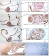
Results.: After three months, granulomas were found in two sheep. In a third sheep we identified a 5 mm diameter cyst in the right lateral ventricle and histological evaluation confirmed that the cyst corresponded to a T. solium larva. Immunohistochemistry with monoclonal antibodies directed against membrane components and excretory/secretory antigens of the T. solium cyst was also used to confirm the etiology of the found granulomas. One of them showed reactivity to the monoclonal antibodies used, thus confirming that it was a cysticercus.
Conclusion.: This experiment is the proof of concept that it is possible to infect sheep with cysticercosis by intracranial inoculation.
Objetivo: . Explorar la viabilidad de desarrollar un modelo de neurocisticercosis (NCC) de oveja mediante infección intracraneal de oncosferas de T. solium.
Materiales y métodos.: Se realizó un modelo de infección experimental de NCC en ovejas. Se inocularon aproximadamente 10 posoncósferas de T. solium cultivadas previamente por 30 días por vía intracraneal en diez ovejas. Las oncósferas, en 0,1 mL de solución salina fisiológica, se inyectaron en el lóbulo parietal a través de una aguja de calibre 18.
Resultados.: Después de tres meses, en dos ovejas se encontraron granulomas y en una tercera identificó un quiste de 5 mm de diámetro en el ventrículo lateral derecho y la evaluación histológica confirmó que el quiste corresponde a una larva de T. solium. También se utilizó inmunohistoquímica con anticuerpos monoclonales dirigidos contra componentes de membrana y antígenos excretorios/secretorios del quiste de T. solium para confirmar la etiología de los granulomas encontrados. Uno de ellos mostro reactividad ante los anticuerpos monoclonales utilizados, confirmando así que se trató de un cisticerco.
Conclusión.: Este experimento es la prueba de concepto de que es posible infectar ovejas con cisticercosis por inoculación intracraneal.
Conflict of interest statement
Figures


References
MeSH terms
Substances
Grants and funding
LinkOut - more resources
Full Text Sources
