

New Contrast Enhancement Method for Multiple Sclerosis Lesion Detection
- PMID: 36478312
- PMCID: PMC10039218
- DOI: 10.1007/s10278-022-00729-1
New Contrast Enhancement Method for Multiple Sclerosis Lesion Detection
Abstract
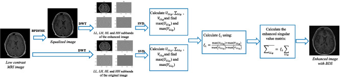
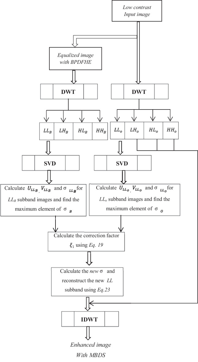
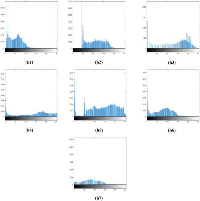
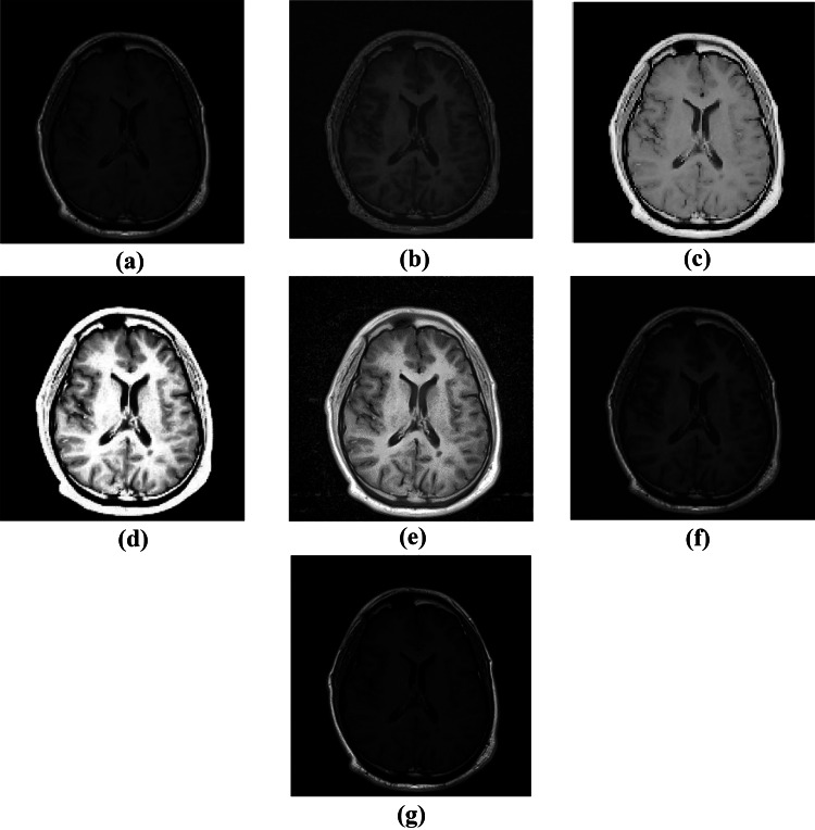
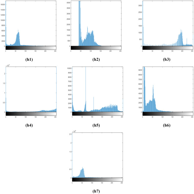
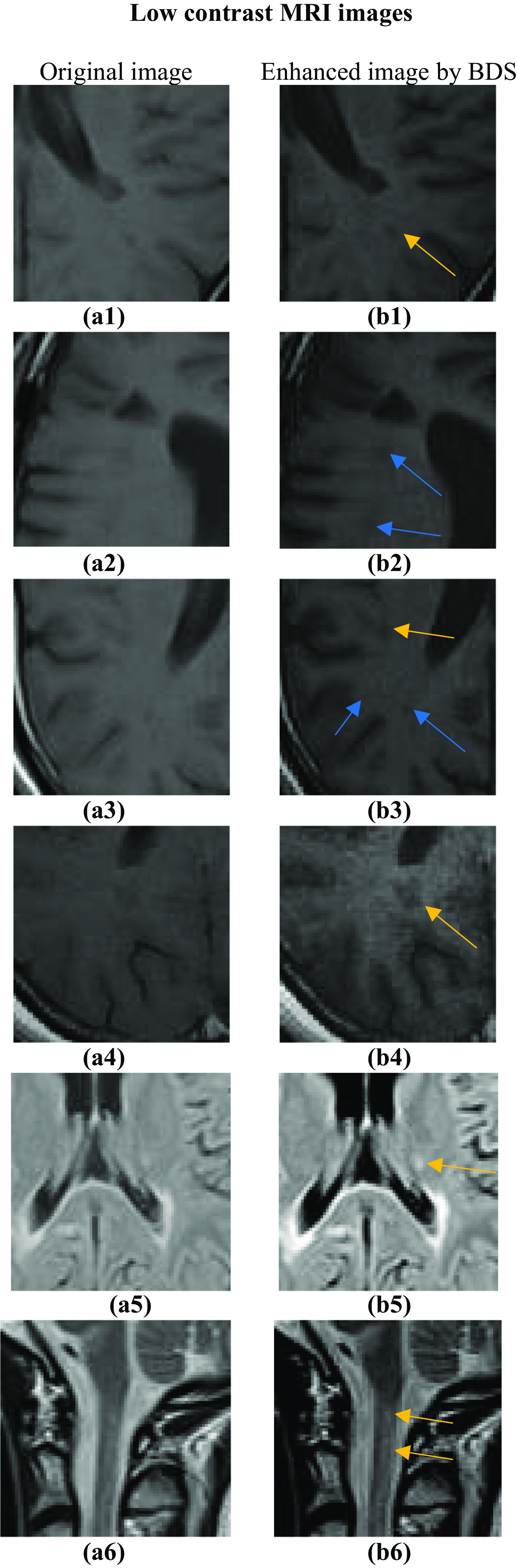
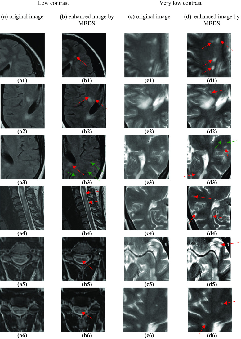
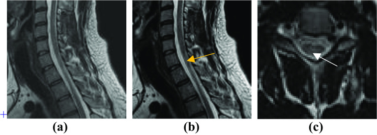
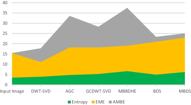
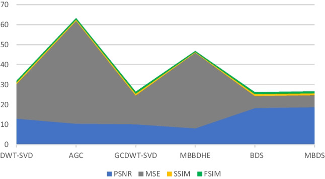
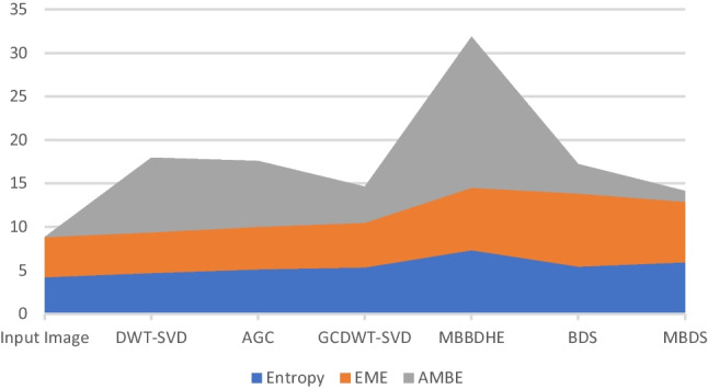
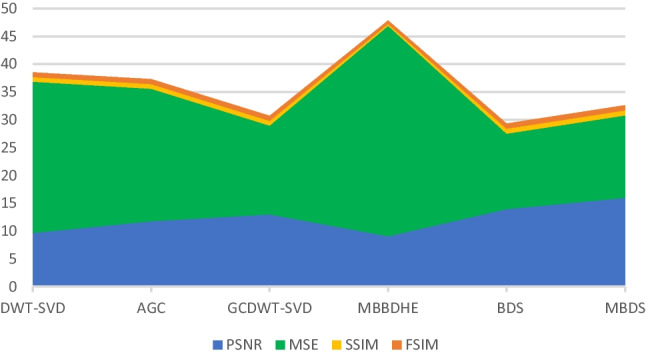
Multiple sclerosis (MS) is one of the most serious neurological diseases. It is the most frequent reason of non-traumatic disability among young adults. MS is an autoimmune disease wherein the central nervous system wrongly destructs the myelin sheath surrounding and protecting axons of nerve cells of the brain and the spinal cord which results in presence of lesions called plaques. The damage of myelin sheath alters the normal transmission of nerve flow at the plaques level, consequently, a loss of communication between the brain and other organs. The consequence of this poor transmission of nerve impulses is the occurrence of various neurological symptoms. MS lesions cause mobility, vision, cognitive, and memory disorders. Indeed, early detection of lesions provides an accurate MS diagnosis. Consequently, and with the adequate treatment, clinicians will be able to deal effectively with the disease and reduce the number of relapses. Therefore, the use of magnetic resonance imaging (MRI) is primordial which is proven as the relevant imaging tool for early diagnosis of MS patients. But, low contrast MRI images can hide important objects in the image such lesions. In this paper, we propose a new automated contrast enhancement (CE) method to ameliorate the low contrast of MRI images for a better enhancement of MS lesions. This step is very important as it helps radiologists in confirming their diagnosis. The developed algorithm called BDS is based on Brightness Preserving Dynamic Fuzzy Histogram Equalization (BPDFHE) and Singular Value Decomposition with Discrete Wavelet Transform (SVD-DWT) techniques. BDS is dedicated to improve the low quality of MRI images with preservation of the brightness level and the edge details from degradation and without added artifacts or noise. These features are essential in CE approaches for a better lesion recognition. A modified version of BDS called MBDS is also implemented in the second part of this paper wherein we have proposed a new method for computing the correction factor. Indeed, with the use of the new correction factor, the entropy has been increased and the contrast is greatly enhanced. MBDS is specially dedicated for very low contrast MRI images. The experimental results proved the effectiveness of developed methods in improving low contrast of MRI images with preservation of brightness level and edge information. Moreover, performances of both proposed BDS and MBDS algorithms exceeded conventional CE methods.
Keywords: BPDFHE; Brightness preservation; Contrast enhancement; Lesion detection; MRI; MS; SVD-DWT.
© 2022. The Author(s) under exclusive licence to Society for Imaging Informatics in Medicine.
Figures
Similar articles
-
A New Adaptive Gamma Correction Based Algorithm Using DWT-SVD for Non-Contrast CT Image Enhancement.IEEE Trans Nanobioscience. 2017 Dec;16(8):666-675. doi: 10.1109/TNB.2017.2771350. IEEE Trans Nanobioscience. 2017. PMID: 29364122
-
An effective method for computerized prediction and segmentation of multiple sclerosis lesions in brain MRI.Comput Methods Programs Biomed. 2017 Mar;140:307-320. doi: 10.1016/j.cmpb.2017.01.003. Epub 2017 Jan 10. Comput Methods Programs Biomed. 2017. PMID: 28254088
-
Correlation between contrast enhanced plaques and plaque diffusion restriction and their signal intensities in FLAIR images in patients who admitted with acute symptoms of multiple sclerosis.J Med Imaging Radiat Sci. 2021 Mar;52(1):121-126. doi: 10.1016/j.jmir.2020.12.001. Epub 2021 Jan 11. J Med Imaging Radiat Sci. 2021. PMID: 33446443
-
The role of magnetic resonance techniques in understanding and managing multiple sclerosis.Brain. 1998 Jan;121 ( Pt 1):3-24. doi: 10.1093/brain/121.1.3. Brain. 1998. PMID: 9549485 Review.
-
Automated detection of multiple sclerosis lesions in serial brain MRI.Neuroradiology. 2012 Aug;54(8):787-807. doi: 10.1007/s00234-011-0992-6. Epub 2011 Dec 20. Neuroradiology. 2012. PMID: 22179659 Review.
Cited by
-
Classification of Lung Diseases Using an Attention-Based Modified DenseNet Model.J Imaging Inform Med. 2024 Aug;37(4):1625-1641. doi: 10.1007/s10278-024-01005-0. Epub 2024 Mar 11. J Imaging Inform Med. 2024. PMID: 38467955 Free PMC article.
References
-
- Thompson AJ, Banwell BL, Barkhof F, Carroll WM, Coetzee T, Comi G, et al.: Diagnosis of multiple sclerosis: 2017 revisions of the McDonald criteria. Lancet Neurol. 17:162–73, 2017. 10.1016/S1474-4422(17)30470-2. - PubMed
-
- Lezak MD, Howieson DB, Bigler ED, Tranel D. Neuropsychological Assessment. 5. New-York: Oxford University Press; 2012.
MeSH terms
LinkOut - more resources
Full Text Sources
Medical
