. 2023 Mar;36(2):232-245.
doi: 10.1111/pcmr.13077. Epub 2023 Jan 3.

Induced pluripotent stem cells reprogramming overcomes technical limitations for highly pigmented adult melanocyte amplification and integration in 3D skin model

Affiliations

Induced pluripotent stem cells reprogramming overcomes technical limitations for highly pigmented adult melanocyte amplification and integration in 3D skin model

Catherine Cohen et al. Pigment Cell Melanoma Res. 2023 Mar.

Abstract

Understanding pigmentation regulations taking into account the original skin color type is important to address pigmentary disorders. Biological models including adult melanocytes from different phenotypes allow to perform fine-tuned explorative studies and support discovery of treatments adapted to populations' skin color. However, technical challenges arise when trying to not only isolate but also amplify melanocytes from highly pigmented adult skin. To bypass the initial isolation and growth of cutaneous melanocytes, we harvested and expanded fibroblasts from light and dark skin donors and reprogrammed them into iPSC, which were then differentiated into melanocytes. The resulting melanocyte populations displayed high purity, genomic stability, and strong proliferative capacity, the latter being a critical parameter for dark skin cells. The iPSC-derived melanocyte strains expressed lineage-specific markers and could be successfully integrated into reconstructed skin equivalent models, revealing pigmentation status according to the native phenotype. In both monolayer cultures and 3D skin models, the induced melanocytes demonstrated responsiveness to promelanogenic stimuli. The data demonstrate that the iPSC-derived melanocytes with high proliferative capacity maintain their pigmentation genotype and phenotypic properties up to a proper integration into 3D skin equivalents, even for highly pigmented cells.

Keywords: induced pluripotent stem cells; lineage specification; melanocyte pigmentation; reconstructed skin model; skin color.


Figures

FIGURE 1
FIGURE 1
Cultivation of adult normal melanocytes from highly pigmented biopsies. Representative proliferation curves for primary normal human melanocyte (NHM) of dark origin in M254 Cascade medium (see section 2 Materials & Methods). Cells obtained from donor D2 (red line) and D3 (blue line) were grown from passage 1 to 5. Dotted lines represent cell growth after a freezing/thawing step (tagged with *).
FIGURE 2
FIGURE 2
Characterization of original skin biopsies. Donor age, ITA° (individual typology angle) value (when available), melanin quantification on Fontana Masson-stained sections are indicated for each donor. Two groups of skin color typologies light and dark were determined according to the above parameters.
FIGURE 3
FIGURE 3
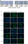
Induced melanocytes from iPSC. (a) Pictures of the six iMc strains cultured in monolayers. Amplification until P6 has been run in parallel. All iMc display a dendritic morphology characteristic of melanocyte cell type, scale bar =100 μm. Respective cell pellets showing melanin content with darker pellets for iMc from darker skins (iMc-D1, iMc-D2, iMc-D3). (b) Characterization of iMc by immunolabeling using antibodies directed against P-Mel17 (HMB45, or NKI-beteb) and TRP1. All iMc are positive for the melanocytic markers, scale bar = 50 μm
FIGURE 4
FIGURE 4
Proliferative activity, cell purity and melanins analysis of iMc strains. (a) Growth curves of the iMc strains. Each strain was sub-cultivated for six passages (P6 to P12). Population doubling (PD) was calculated as follows: LOG10 (harvested cells number/seeded cells number) / LOG10(2). PD was obtained for each passage and cumulated PD was plotted along time for each iMc strain. Pictures of cell pellets obtained at passage 12 showing darker pellets for iMc from dark skin donors. (b) Cytometric analysis of melanocytic proteins P-Mel17 (HMB45 and NKI-beteb) and TRP1. Notice the high purity level of all iMc strains. Illustration of P-Mel17 (NKI-beteb) FACS plots is shown for iMc-L3 and iMc-D3. (c) Melanins analysis of iMc strains in parallel with two NHM neonatal reference strains showed results for Total melanin (TM expressed in μg/million cells) by spectrophotometry or HPLC and the linear correlation between them. The strains were submitted to HPLC dosages (expressed in ng/million cells) for PTCA and PDCA (reflecting DHICA and DHI moieties) from eumelanin and TTCA (reflecting benzothiazole moiety) from phaeomelanin. The dosages of these markers were coherent with the phenotypical groups.
FIGURE 5
FIGURE 5
Characterization of PRS model after iMc integration. Representative results from one experiment where all iMc strains were performed in parallel. Results were replicated within a total of three independent experiments. Histology quality control after HES staining: All samples displayed a correct 3D organization with fully differentiated epidermis and stratum corneum (pink), scale bar = 50 μm. Fontana Masson staining of skin sections reveals melanin as black dots quantified by the ratio of the surface covered by melanin over the epidermal surface and expressed by mean ± SD of the triplicate samples. Higher magnification shows melanocytes with dendrites and melanin pigment within epidermal keratinocytes, scale bar = 50 μm. TRP1 immunostaining on epidermal sheets highlights melanocytes (green cells) homogeneously distributed at the basal layer, scale bar = 100 μm. Pictures indicate the iMc counting values as mean ± SD cell/mm2 calculated from each triplicate sample. DOPA reaction reveals more intense DOPA activity for cells of dark origin, scale bar = 50 μm. Immunostaining of tyrosinase on skin sections outlines in red the iMc localized at the basal layer of the epidermis, scale bar = 50 μm. Results were confirmed in other experiments (n = 3 for each strain).
FIGURE 6
FIGURE 6
Integration of iMc, neonatal primary melanocyte and adult primary melanocyte from dark origin in 3D skin model. Illustration of comparative integration of iMc-D3 (induced melanocyte from D3 adult biopsy), of neonatal NHM (a primary melanocyte from dark origin) and NHM-D3 (adult primary melanocyte from D3 biopsy). HES staining displays the correct skin reconstruction. Fontana Masson staining labels melanin production. Measurement of melanin density was calculated by ratio of the surface covered by melanin over the epidermal surface. DOPA reaction illustrates the strong DOPA activity of melanocytes. TRP1 labeling at the dermal epidermal junction of PRS enables melanocyte (green) counts per mm2. Notice the very low amount of NHM-D3 integrated in PRS. All conditions were performed in triplicates and numbers indicate means and standard deviations.
FIGURE 7
FIGURE 7
Functionality of iMc strains under melanogenic stimulation. Representative results from one experiment where all iMc strains were performed in parallel. Results were replicated in two independent experiments. (a) iMc strains were incubated for 3 days in PDBU supplemented culture medium before stimulation with 10 μM Forskolin for 48 h. After cell lysis, centrifugation, melanin pellet solubilization in NaOH 1 N, samples were deposited in a microplate for optical density (OD) reading at 405 nm. (b) Graph shows resulting melanin concentration means ± SD for iMc-L and iMc-D groups stimulated by Forskolin versus control (each group includes 3 donors × 2 replicates). Significant differences are indicated between conditions (*p < .05). (c) Macroscopic view of triplicate PRS samples under basal (ctrl) or stimulated conditions by αMSH 50 nM or fibroblast deletion (w/o fib), scale bar = 15 mm. (d) Graph of corresponding spectrocolorimetric luminance values expressed as mean value ± SD from each donor’s PRS triplicates. (e) Graph of luminance values expressed as mean values ± SD of samples from light (iMc-L) or dark (iMc-D) origin (each group includes 3 donors × 3 PRS replicates). (f) Graph of melanin density quantified on PRS sections stained by Fontana Masson, expressed as mean ± SD for each iMc strain condition. (g) Graph of melanin density expressed as mean value ± SD of samples from light (iMc-L) or dark (iMc-D) origin (each group includes 3 donors × 3 PRS replicates). (h) Pictures from representative Fontana Masson staining of PRS samples from light (L3) or dark (D3) origin. Melanin contents (black dots) are increased after melanogenesis stimulation, scale bar = 50 μm.

Similar articles

Cited by

References

    1. Abdel-Malek ZA, Swope VB, Nordlund JJ, & Medrano EE (1994). Proliferation and propagation of human melanocytes in vitro are affected by donor age and anatomical site. Pigment Cell Research, 7(2), 116–122. 10.1111/j.1600-0749.1994.tb00035.x - DOI - PubMed
    1. Attwood SW, & Edel MJ (2019). iPS-cell technology and the problem of genetic instability-can it ever Be safe for clinical use? Journal of Clinical Medicine, 8(3), 288. 10.3390/jcm8030288 - DOI - PMC - PubMed
    1. Bandyopadhyay D, & Medrano EE (2000). Melanin accumulation accelerates melanocyte senescence by a mechanism involving p16INK4a/CDK4/pRB and E2F1. Annals of the New York Academy of Sciences, 908, 71–84. 10.1111/j.1749-6632.2000.tb06637.x - DOI - PubMed
    1. Boyce ST, Lloyd CM, Kleiner MC, Swope VB, Abdel-Malek Z, & Supp DM (2017). Restoration of cutaneous pigmentation by transplantation to mice of isogeneic human melanocytes in dermal-epidermal engineered skin substitutes. Pigment Cell & Melanoma Research, 30(6), 531–540. 10.1111/pcmr.12609 - DOI - PubMed
    1. Chardon A, Cretois I, & Hourseau C (1991). Skin colour typology and suntanning pathways. International Journal of Cosmetic Science, 13(4), 191–208. 10.1111/j.1467-2494.1991.tb00561.x - DOI - PubMed

Publication types