

Plasma inflammatory biomarkers in cerebral small vessel disease: A review
- PMID: 36478511
- PMCID: PMC9873530
- DOI: 10.1111/cns.14047
Plasma inflammatory biomarkers in cerebral small vessel disease: A review
Abstract
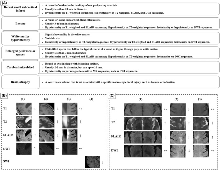
Cerebral small vessel disease (CSVD) is a group of pathological processes affecting small arteries, arterioles, capillaries, and small veins of the brain. It is one of the most common subtypes of cerebrovascular diseases, especially highly prevalent in elderly populations, and is associated with stroke occurrence and recurrence, cognitive impairment, gait disorders, psychological disturbance, and dysuria. Its diagnosis mainly depends on MRI, characterized by recent small subcortical infarcts, lacunes, white matter hyperintensities (WMHs), enlarged perivascular spaces (EPVS), cerebral microbleeds (CMBs), and brain atrophy. While the pathophysiological processes of CSVD are not fully understood at present, inflammation is noticed as playing an important role. Herein, we aimed to review the relationship between plasma inflammatory biomarkers and the MRI features of CSVD, to provide background for further research.
Keywords: cerebral small vessel disease; endothelial dysfunction; imaging features; inflammatory biomarkers.
© 2022 The Authors. CNS Neuroscience & Therapeutics published by John Wiley & Sons Ltd.
Conflict of interest statement
The authors declare that this work has no conflict of interest.
Figures
Similar articles
-
Cerebral small vessel disease and post-stroke cognitive impairment.Int J Neurosci. 2017 Sep;127(9):824-830. doi: 10.1080/00207454.2016.1261291. Epub 2016 Nov 28. Int J Neurosci. 2017. PMID: 27838946 Review.
-
Cerebral Small Vessel Disease.Cell Transplant. 2018 Dec;27(12):1711-1722. doi: 10.1177/0963689718795148. Epub 2018 Sep 25. Cell Transplant. 2018. PMID: 30251566 Free PMC article. Review.
-
Correlations of Plasma Biomarkers and Imaging Characteristics of Cerebral Small Vessel Disease.Brain Sci. 2024 Mar 12;14(3):269. doi: 10.3390/brainsci14030269. Brain Sci. 2024. PMID: 38539657 Free PMC article. Review.
-
Association of inflammatory markers with cerebral small vessel disease in community-based population.J Neuroinflammation. 2022 May 6;19(1):106. doi: 10.1186/s12974-022-02468-0. J Neuroinflammation. 2022. PMID: 35513834 Free PMC article.
-
Association between remote diffusion-weighted imaging lesions and cerebral small vessel disease in primary intracerebral hemorrhage.Eur J Neurol. 2019 Jul;26(7):961-968. doi: 10.1111/ene.13915. Epub 2019 Mar 5. Eur J Neurol. 2019. PMID: 30742740
Cited by
-
Novel inflammatory and insulin resistance indices provide a clue in cerebral amyloid angiopathy.Sci Rep. 2024 May 20;14(1):11474. doi: 10.1038/s41598-024-62280-z. Sci Rep. 2024. PMID: 38769356 Free PMC article.
-
Proteogenomics in cerebrospinal fluid and plasma reveals new biological fingerprint of cerebral small vessel disease.Res Sq [Preprint]. 2024 Jul 2:rs.3.rs-4535534. doi: 10.21203/rs.3.rs-4535534/v1. Res Sq. 2024. PMID: 39011113 Free PMC article. Preprint.
-
Cerebral Small Vessel Disease: Therapeutic Approaches Targeting Neuroinflammation, Oxidative Stress, and Endothelial Dysfunction.Curr Issues Mol Biol. 2025 Mar 27;47(4):232. doi: 10.3390/cimb47040232. Curr Issues Mol Biol. 2025. PMID: 40699631 Free PMC article. Review.
-
Causal relationship of inflammatory cytokines and serum metabolites in cerebral small vessel disease: a two-step Mendelian randomization study.Eur J Neurol. 2024 Dec;31(12):e16443. doi: 10.1111/ene.16443. Epub 2024 Aug 16. Eur J Neurol. 2024. PMID: 39150083 Free PMC article.
-
Value of blood neural cell-derived small extracellular vesicles in the diagnosis and prediction of Alzheimer's disease: A systematic review.J Prev Alzheimers Dis. 2025 Aug;12(7):100193. doi: 10.1016/j.tjpad.2025.100193. Epub 2025 May 1. J Prev Alzheimers Dis. 2025. PMID: 40316481 Free PMC article.
References
-
- Pantoni L. Cerebral small vessel disease: from pathogenesis and clinical characteristics to therapeutic challenges. Lancet Neurol. 2010;9:689‐701. - PubMed
-
- Staszewski J, Skrobowska E, Piusińska‐Macoch R, Brodacki B, Stępień A. IL‐1α and IL‐6 predict vascular events or death in patients with cerebral small vessel disease‐data from the SHEF‐CSVD study. Adv Med Sci. 2019;64:258‐266. - PubMed
-
- Conijn MM, Kloppenborg RP, Algra A, et al. Cerebral small vessel disease and risk of death, ischemic stroke, and cardiac complications in patients with atherosclerotic disease: the second manifestations of ARTerial disease‐magnetic resonance (SMART‐MR) study. Stroke. 2011;42:3105‐3109. - PubMed
Publication types
MeSH terms
Substances
LinkOut - more resources
Full Text Sources
Medical
