Review
. 2022 Nov 21:10:1060850.
doi: 10.3389/fcell.2022.1060850. eCollection 2022.

Unfolding the role of placental-derived Extracellular Vesicles in Pregnancy: From homeostasis to pathophysiology

Affiliations
Review

Unfolding the role of placental-derived Extracellular Vesicles in Pregnancy: From homeostasis to pathophysiology

Miguel A Ortega et al. Front Cell Dev Biol. .

Abstract

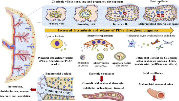
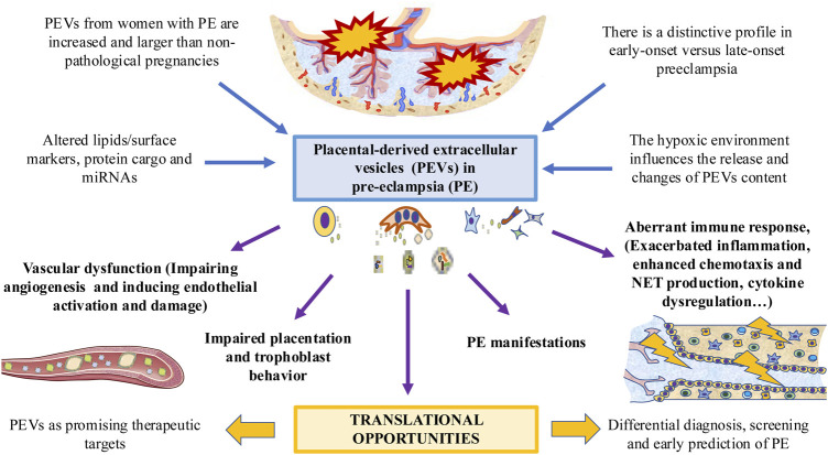
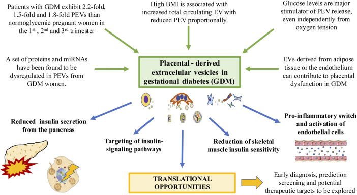
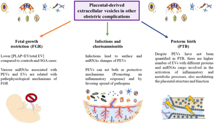
The human placenta is a critical structure with multiple roles in pregnancy, including fetal nutrition and support, immunological, mechanical and chemical barrier as well as an endocrine activity. Besides, a growing body of evidence highlight the relevance of this organ on the maternofetal wellbeing not only during gestation, but also from birth onwards. Extracellular vesicles (EVs) are complex macromolecular structures of different size and content, acting as carriers of a diverse set of molecules and information from donor to recipient cells. Since its early development, the production and function of placental-derived EVs are essential to ensure an adequate progress of pregnancy. In turn, the fetus receives and produce their own EVs, highlighting the importance of these components in the maternofetal communication. Moreover, several studies have shown the clinical relevance of EVs in different obstetric pathologies such as preeclampsia, infectious diseases or gestational diabetes, among others, suggesting that they could be used as pathophysiological biomarkers of these diseases. Overall, the aim of this article is to present an updated review of the published basic and translational knowledge focusing on the role of placental-derived EVs in normal and pathological pregnancies. We suggest as well future lines of research to take in this novel and promising field.

Keywords: decidualization; extracellular vesicles; gestational diabetes mellitus; normal pregnancy; obstetric complications; placental-derived extracellular vesicles; placentation; pre-eclampsia.


Conflict of interest statement

The authors declare that the research was conducted in the absence of any commercial or financial relationships that could be construed as a potential conflict of interest.

Figures

FIGURE 1
FIGURE 1
A general overview of PEVs. Biosynthesis and release of PEVs increase as pregnancy progresses along with the development of the placental structures. Syncytiotrophoblasts, which are in contact with maternal blood (Intervillous space), are a major source of PEVs, although cytotrophoblast, Hofbauer cells, mesenchymal cells, extravillous trophoblasts and others also produce them. PEVs are essential for the interplay between the placenta and endometrial decidua, modulating both the placentation and decidualization processes. PEVs also modulate the immune system, the inflammatory response and immune tolerance. PEVs reach systemic circulation or enter through fetal capillaries into the fetal circulation, playing a key role in the crosstalk between the placenta with maternal and fetal tissues.
FIGURE 2
FIGURE 2
The role of PEVs in PE. PEVs are increased in women affected with this condition, with multiple changes in their surface and cargo. These changes are different in early versus late-onset PE. A possible modulator of PEVs is the low oxygen tension and hypoxic environment. PEVs released by the placenta participates in the PE-associated vascular dysfunction, impairing placentation and trophoblast behavior, dysregulating the immune system and leading to different PE manifestations. This can arise potential translational opportunities for exploring.
FIGURE 3
FIGURE 3
The relevance of PEVs in the context of GDM. Different factors associated with this condition such as hyperglycemia and high body mass index can induce significant changes in PEV profile. EVs from other tissues affected by GDM such as adipose tissue can induce changes in the placenta. PEVs can in turn reduce insulin secretion from the pancreas, altering insulin signaling pathways and insulin sensitivity in different tissues such as the muscle or endothelium, driving to its activation and pro-inflammatory switch. Translational knowledge will help to improve the clinical management of GDM patients.
FIGURE 4
FIGURE 4
A graphical abstract of some of the current notions of PEVs in different obstetric pathologies. As represented, the role of PEVs remains to be further explored in the field of fetal growth restriction, infections and preterm birth. However, some initial results are starting to arise in these conditions, aiding to improve our knowledge of these obstetric complications. Due to the relevance of PEVs in many physiological and pathological processes, we encourage for future studies in these areas.

Similar articles

Cited by

References

    1. Aaltonen R., Heikkinen T., Hakala K., Laine K., Alanen A. (2005). Transfer of proinflammatory cytokines across term placenta. Obstet. Gynecol. 106, 802–807. 10.1097/01.AOG.0000178750.84837.ed - DOI - PubMed
    1. Aharon A., Katzenell S., Tamari T., Brenner B. (2009). Microparticles bearing tissue factor and tissue factor pathway inhibitor in gestational vascular complications. J. Thromb. Haemost. 7, 1047–1050. 10.1111/J.1538-7836.2009.03342.X - DOI - PubMed
    1. Akers J. C., Gonda D., Kim R., Carter B. S., Chen C. C. (2013). Biogenesis of extracellular vesicles (EV): Exosomes, microvesicles, retrovirus-like vesicles, and apoptotic bodies. J. Neurooncol. 113, 1–11. 10.1007/S11060-013-1084-8 - DOI - PMC - PubMed
    1. Alfadhli E. M. (2015). Gestational diabetes mellitus. Saudi Med. J. 36, 399–406. 10.15537/SMJ.2015.4.10307 - DOI - PMC - PubMed
    1. Aplin J. D., Lewis R. M., Jones C. J. P. (2018). Development of the human placental villus. Ref. Modul. Biomed. Sci. 1, 1. 10.1016/B978-0-12-801238-3.99857-X - DOI

LinkOut - more resources