. 2022 Nov 28;8(12):e11778.
doi: 10.1016/j.heliyon.2022.e11778. eCollection 2022 Dec.

Multispectral imaging for MicroChip electrophoresis enables point-of-care newborn hemoglobin variant screening

Affiliations

Multispectral imaging for MicroChip electrophoresis enables point-of-care newborn hemoglobin variant screening

Ran An et al. Heliyon. .

Abstract

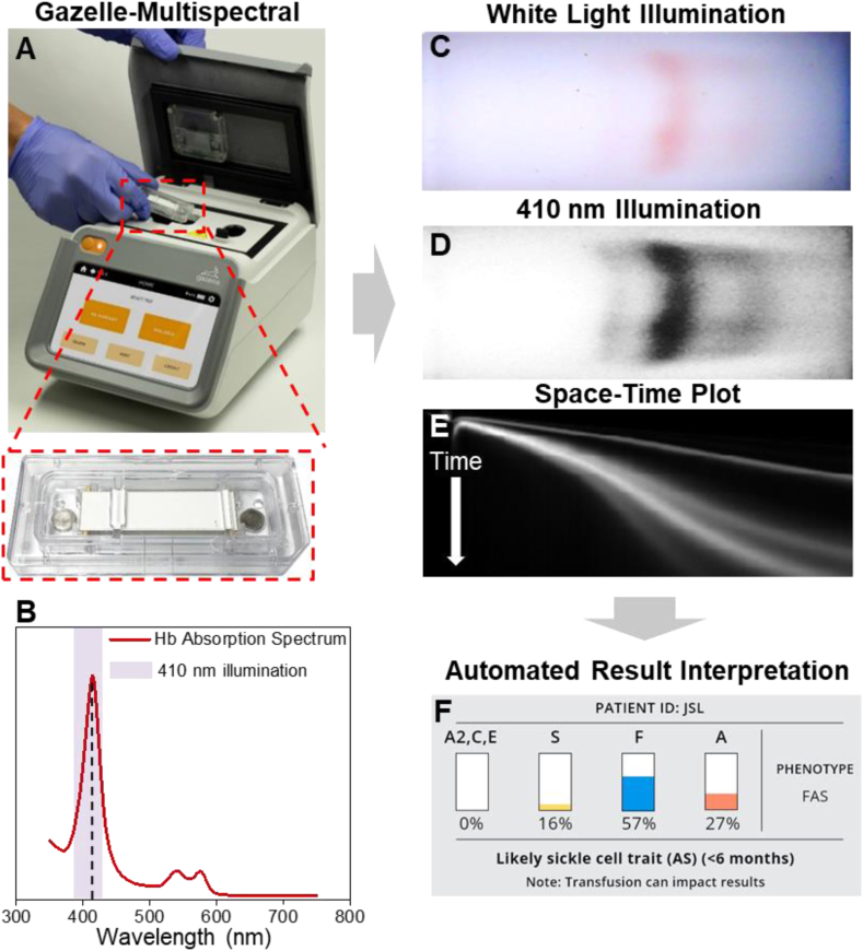
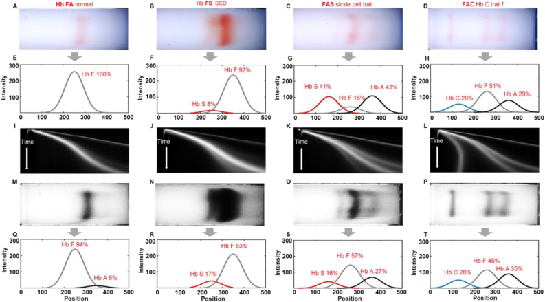
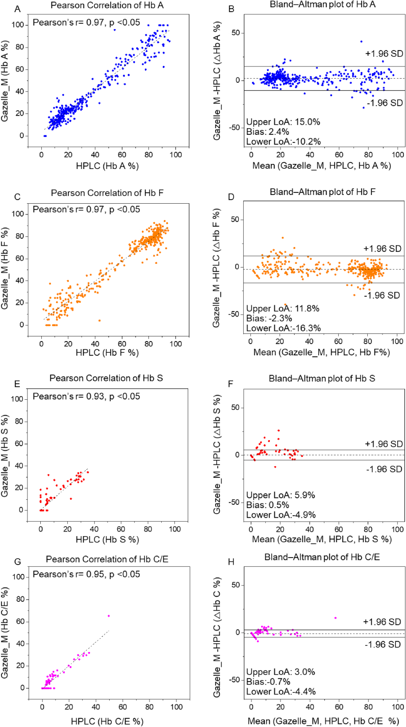
Hemoglobin (Hb) disorders affect nearly 7% of the world's population. Globally, around 400,000 babies are born annually with sickle cell disease (SCD), primarily in sub-Saharan Africa where morbidity and mortality rates are high. Screening, early diagnosis, and monitoring are not widely accessible due to technical challenges and cost. We hypothesized that multispectral imaging will allow sensitive hemoglobin variant identification in existing affordable paper-based Hb electrophoresis. To test this hypothesis, we developed the first integrated point-of-care multispectral Hb variant test: Gazelle-Multispectral. Here, we evaluated the accuracy of Gazelle-Multispectral for Hb variant newborn screening in 265 newborns with known hemoglobin variants including hemoglobin A (Hb A), hemoglobin F (Hb F), hemoglobin S (Hb S) and hemoglobin C (Hb C). Gazelle-Multispectral detected levels of Hb A, Hb F, Hb S, and Hb C/E/A2, demonstrated high correlations with the results reported by laboratory gold standard high performance liquid chromatography (HPLC) at Pearson Correlation Coefficient = 0.97, 0.97, 0.93, and 0.95. Gazelle-Multispectral demonstrated accuracy of 96.8% in subjects of 0-3 days, and 96.9% in newborns. The ability to obtain accurate results on newborn samples suggest that Gazelle-Multispectral can be suitable for large-scale newborn screening and for diagnosis of SCD in low resource settings.

Keywords: Genetic hemoglobin disorders; Multispectral imaging; Newborn screening; Point-of-care diagnostics; Sickle cell disease.


Conflict of interest statement

The authors declare the following conflict of interests: RA, QZ, UAG, and Case Western Reserve University have financial interests in Hemex Health Inc. UAG and Case Western Reserve University have financial interests in BioChip Labs Inc. UAG and Case Western Reserve University have financial interests in Xatek Inc. UAG has financial interests in DxNow Inc. Financial interests include licensed intellectual property, stock ownership, research funding, employment, and consulting. Hemex Health Inc. offers point-of-care diagnostics for hemoglobin disorders, anemia, and malaria. BioChip Labs Inc. offers commercial clinical microfluidic biomarker assays for inherited or acquired blood disorder.

Figures

Figure 1
Figure 1
Gazelle-Multispectral for screening hemoglobin variants in newborns. (A) Gazelle-Multispectral platform for paper-based microchip electrophoresis using disposable cartridge (red box) at the point of need. (B) Hemoglobin absorption spectrum. Purple shaded area covers more than 90% of the power output according to the LED manufacturing specification and our internal testing. The Gazelle-Multispectral perform real time imaging and data analysis tracking the Hb electrophoresis process under both white light illumination (C) and 410 nm illumination (D). The images captured under white light illumination provides visual validation of test progression. (E) The space-time plots generated based on the images captures under 410 nm illumination are used for identifying and quantifying Hb variants in real time using an internally integrated data analysis algorithm. (F) At the end of each test, Gazelle-Multispectral algorithm automatically reports the identification and quantification of Hb variant results, and determines the patient phenotype accordingly.
Figure 2
Figure 2
Identification of Hb variants and quantification of Hb percentages by Gazelle-Multispectral. (A-D) The first row shows images captured under white light field. (E-H) The second row shows electropherograms reconstructed by the white data analysis algorithm based on the white light images (electropherograms not visible by users in the field). (I-L) The third row illustrates 2D representation of Gazelle-Multispectral space-time plots of band migration in 410 nm imaging mode, which are used with the machine learning algorithm for identifying Hb variants. (M-P) The fourth row shows images captured under 410 nm wavelength. (Q-T) The fifth row shows electropherograms reconstructed by the data analysis algorithm based on the 410 nm images captured at the same time as the white light images. The Gazelle-Multispectral data analysis algorithm sensitively and accurately identified Hb variants agreeing with HPLC reported results. Column 1–4: Multispectral test results for samples with different phenotypes. Column 1: Hb FA (Healthy newborn, Hb A: 8% vs. 6%, Hb F: 92% vs. 94%, Gazelle-Multispectral vs. HPLC); Column 2: Hb FS (Newborn with sickle cell disease, Hb F 83% vs. 89%, Hb S 17% vs. 11%, Gazelle-Multispectral vs. HPLC); Column 3: Hb FAS (Newborn with sickle cell trait, Hb S 16% vs. 16%, Hb F 57% vs. 55%, Hb A 27% vs. 29%, Gazelle-Multispectral vs. HPLC); and Column 4: Hb FAC (Newborn with Hb C disease, Hb C 20% vs. 20%, Hb F 45% vs. 45%, Hb A 35% vs. 35%, Gazelle-Multispectral vs. HPLC). Gazelle-Multispectral enabled identification and quantification of low concentration Hb variants with higher sensitivity (I–T) compared to white light imaging mode (A-H). †: Gazelle-Multispectral reports Hb C/E/A2 as demonstrated in Figure 1E. The identified Hb C/E/A2 were recognized as Hb C according to test location (Ghana).
Figure 3
Figure 3
Gazelle-Multispectral Hb variant identification and quantification in all test subjects. Pearson correlation (Column 1) and Bland-Altman analysis (Column 2) showed Gazelle-Multispectral identified and quantified Hb A (A&B, Pearson coefficient correlation (PCC) = 0.97, p < 0.05, Mean bias ±1.96 × Standard Deviation (SD) = 2.4% ± 12.6%), Hb F (C&D, PCC = 0.97, p < 0.05, Mean bias ±1.96SD = −2.3% ± −14.0%), Hb S (E&F, PCC = 0.93, p < 0.05, Mean bias ±1.96 SD = 0.5% ± 5.4%), and Hb C levels (G&H, PCC = 0.95, p < 0.05, Mean bias ±1.96SD = −0.7% ± 3.3%) agree with the ones reported by laboratory standard HPLC. In Column 2, the solid black lines indicate the mean biases and the dashed gray lines represent 95% limits of agreement. ∗: 365 ‘Valid’ tests out of 441 total tests were included in this correlation calculation. ‘Inconclusive’ tests did not generate a result that could be included in the correlation coefficient calculation [39, 40].

Similar articles

  • Point-of-Care Diagnostic Test for Beta-Thalassemia.
    An R, Avanaki A, Thota P, Nemade S, Mehta A, Gurkan UA. An R, et al. Biosensors (Basel). 2024 Feb 2;14(2):83. doi: 10.3390/bios14020083. Biosensors (Basel). 2024. PMID: 38392002 Free PMC article.
  • Evaluation of Microchip-Based Point-Of-Care Device "Gazelle" for Diagnosis of Sickle Cell Disease in India.
    Shrivas S, Patel M, Kumar R, Gwal A, Uikey R, Tiwari SK, Verma AK, Thota P, Das A, Bharti PK, Shanmugam R. Shrivas S, et al. Front Med (Lausanne). 2021 Oct 13;8:639208. doi: 10.3389/fmed.2021.639208. eCollection 2021. Front Med (Lausanne). 2021. PMID: 34722554 Free PMC article.
  • Paper-based microchip electrophoresis for point-of-care hemoglobin testing.
    Hasan MN , Fraiwan A , An R , Alapan Y , Ung R , Akkus A , Xu JZ , Rezac AJ , Kocmich NJ , Creary MS , Oginni T , Olanipekun GM , Hassan-Hanga F , Jibir BW , Gambo S , Verma AK , Bharti PK , Riolueang S , Ngimhung T , Suksangpleng T , Thota P , Werner G , Shanmugam R , Das A , Viprakasit V , Piccone CM , Little JA , Obaro SK , Gurkan UA . Hasan MN , et al. Analyst. 2020 Apr 7;145(7):2525-2542. doi: 10.1039/c9an02250c. Epub 2020 Mar 3. Analyst. 2020. PMID: 32123889 Free PMC article.
  • Hemoglobin e syndromes.
    Vichinsky E. Vichinsky E. Hematology Am Soc Hematol Educ Program. 2007:79-83. doi: 10.1182/asheducation-2007.1.79. Hematology Am Soc Hematol Educ Program. 2007. PMID: 18024613 Review.
  • Carrier screening for thalassemia and hemoglobinopathies in Canada.
    Langlois S, Ford JC, Chitayat D; CCMG PRENATAL DIAGNOSIS COMMITTEE; SOGC GENETICS COMMITTEE. Langlois S, et al. J Obstet Gynaecol Can. 2008 Oct;30(10):950-959. doi: 10.1016/S1701-2163(16)32975-9. J Obstet Gynaecol Can. 2008. PMID: 19038079 Review. English, French.

Cited by

References

    1. The World Health Organization . 2022. Newborn Health in the Western Pacific.https://www.who.int/westernpacific/health-topics/newborn-health [cited 2022 January, 11]; Available from:
    1. Alapan Y., Fraiwan A., Kucukal E., Hasan M.N., Ung R., Kim M., Odame I., Little J.A., Gurkan U.A. Emerging point-of-care technologies for sickle cell disease screening and monitoring. Expet Rev. Med. Dev. 2016;13(12):1073–1093. - PMC - PubMed
    1. McGann Patrick T., Hoppe Carolyn. The pressing need for point-of-care diagnostics for sickle cell disease: a review of current and future technologies. Blood Cells, Mol. Dis. 2017;67:104–113. - PMC - PubMed
    1. Hsu L., Nnodu O.E., Brown B.J., Tluway F., King S., Dogara L.G., Patil C., Shevkoplyas S.S., Lettre G., Cooper R.S., Gordeuk V.R., Tayo B.O. White paper: pathways to progress in newborn screening for sickle cell disease in sub-Saharan Africa. J. Trop. Dis. Publ. Health. 2018;6(2):260. - PMC - PubMed
    1. El-Haj Nura, Hoppe Carolyn C. Newborn screening for SCD in the USA and Canada. Int. J. Neonatal Screen. 2018;4(4):36. - PMC - PubMed

LinkOut - more resources