. 2022 Dec 1:33:101399.
doi: 10.1016/j.bbrep.2022.101399. eCollection 2023 Mar.

X-ray-irradiated K562 feeder cells for expansion of functional CAR-T cells

Affiliations

X-ray-irradiated K562 feeder cells for expansion of functional CAR-T cells

Khac Cuong Bui et al. Biochem Biophys Rep. .

Abstract

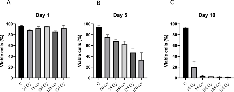
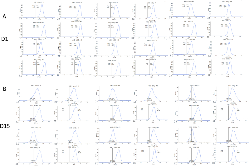
Immunotherapy, particularly CAR-T therapy has recently emerged as an innovator for cancer treatment. Gamma-irradiated K562 cells is a common and effective method to stimulated CAR-T cells prior to treatment. However, high cost and limited equipment of gamma-irradiation is drawback of this method. This requires the establishment of CAR-T-expanding alternatives, such as X-ray-irradiated K562 cells. X-ray irradiation was used to deactivate K562 cells. The post-irradiative cell survival was investigated by counting of the number of cells, staining with Trypan Blue and PI. FACS analysis was applied to detect the expression of cell surface markers. The production of CD19-CAR-T cells were executed from fresh blood donor by CD19-CAR-plasmid transfection, followed by the stimulation with X-ray-irradiated K562 feeder cells. The function of produced CAR-T cells was checked by their ability to kill Daudi cells. X-ray-irradiation inhibited the propagation and viability of K562 cells in a dose- and time-dependent manner. Interestingly, CAR-T-stimulating effectors were remained on the surface of X-ray-irradiated K562 cells. CD-19-CAR-T cells were produced successfully, suggested by number of CAR-positive cells in transfected and stimulated population, compared to un-transfected group. Lastly, our data showed that engineered CAR-T cells effectively killed Daudi cells. Our data demonstrated the efficacy of X-ray on deactivation K562 feeder cells which subsequently stimulated and expanded functional CAR-T cells. Thus, X-ray can be used as an alternative to inactivate K562 cells prior to using as a feeder of CAR-T cells.

Keywords: CAR-T immunotherapy; K562 feeder cells; X-ray irradiation.


Conflict of interest statement

The authors declare no potential conflicts of interest.

Figures

Fig. 1
Fig. 1
X-ray irradiation effectively inhibits the proliferation of K562 cells. K562 cells were plated at the concentration of 105 cells/well for 24 h before irradiated with different doses of X-ray (0, 50, 75, 100, 125, 150 Gy). The cell number of irradiated K562 cells were counted on 1-, 5-, 10-day post-irradiation. (A) The number of irradiated K562 cells on day one. (B) The number of irradiated K562 cells on day five. (C) The number of irradiated K562 cells on day ten..
Fig. 2
Fig. 2
The post-irradiative viability of K562-aAPC cells. K562 cells were plated at the concentration of 105 cells/well for 24 h before irradiated with different doses of X-ray (0, 50, 75, 100, 125, 150 Gy). Then, the cells were stained with Trypan Blue on day one, five, ten of post-irradiation. (A) The viability of irradiated K562 cells on day one. (B) The viability of irradiated K562 cells on day five. (C) The viability of irradiated K562 cells on day ten.
Fig. 3
Fig. 3
CAR-T-stimulating markers are remained on irradiated K562 cells. K562 cells were plated at the concentration of 105 cells/well for 24 h before irradiated with different doses of X-ray (0, 50, 75, 100, 125, 150 Gy). Then, the cells were prepared and analysed for FACS assay detecting interesting surface markers, including CD19 (FITC), CD64 (APC-Cy7), CD86 (APC) and CD137L (PE) after one day (A) and 15 days (B) of post-irradiation.
Fig. 4
Fig. 4
Effective production of CAR-T cells by CD19-plasmid transfection, followed by co-culture with K562 cells. In-laboratory produced CAR-T cells effectively kill Daudi cells. PBMCs were freshly isolated from blood of healthy blood donors using density gradient centrifugation, and K562-aAPC cells were cultured and transfected with CD19, CD64, CD86, and CD137L. Afterwards, freshly prepared PBMCs were transfected with designated CoOppSBi/CD19RCD137 plus pCMV(CAT)T7-SB100, followed by co-culturing with 100 Gy X-ray-irradiated K562-aAPC cells. The expression of CD8+-CAR+-T was detected by FACS. (A) The expression of CAR+ on PBMCs population; (B) The expression of CAR+ on tranfected PBMCs which co-cultured with 100 Gy X-ray-irradiated K562-aAPC cells. In-laboratory produced CAR-T cells effectively kill Daudi cells. Daudi cells were cultured and seeded in culturing medium at the density of 105 cells/six-well plate for 24 h before being stained with CFSE for 15 min. Afterwards, CFSE-stained Daudi cells were co-cultured with transfected and stimulated CAR-T population or PBMCs, and viability of Daudi cells were determined by FACS analysis detecting 7-AAD staining. (C) Daudi cells co-cultured with PBMCs. (D) Daudi cells co-cultured with CAR-T cells.

References

    1. Bray F., et al. Global cancer statistics 2018: GLOBOCAN estimates of incidence and mortality worldwide for 36 cancers in 185 countries. CA A Cancer J. Clin. 2018;68(6):394–424. - PubMed
    1. Ferlay J., et al. Cancer incidence and mortality worldwide: sources, methods and major patterns in GLOBOCAN 2012. Int. J. Cancer. 2015;136(5):E359–E386. - PubMed
    1. Maude S.L., et al. Chimeric antigen receptor T cells for sustained remissions in leukemia. N. Engl. J. Med. 2014;371(16):1507–1517. - PMC - PubMed
    1. Brentjens R.J., et al. CD19-targeted T cells rapidly induce molecular remissions in adults with chemotherapy-refractory acute lymphoblastic leukemia. Sci. Transl. Med. 2013;5(177) - PMC - PubMed
    1. Lee D.W., et al. T cells expressing CD19 chimeric antigen receptors for acute lymphoblastic leukaemia in children and young adults: a phase 1 dose-escalation trial. Lancet. 2015;385(9967):517–528. - PMC - PubMed