

Effect of integrin α7 on cell proliferation, invasion, apoptosis and the PI3K/AKT pathway, and its association with clinicopathological features in endometrial cancer
- PMID: 36478908
- PMCID: PMC9713827
- DOI: 10.3892/ol.2022.13612
Effect of integrin α7 on cell proliferation, invasion, apoptosis and the PI3K/AKT pathway, and its association with clinicopathological features in endometrial cancer
Abstract
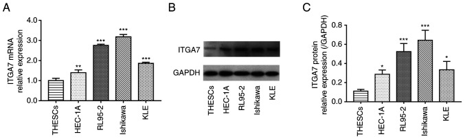
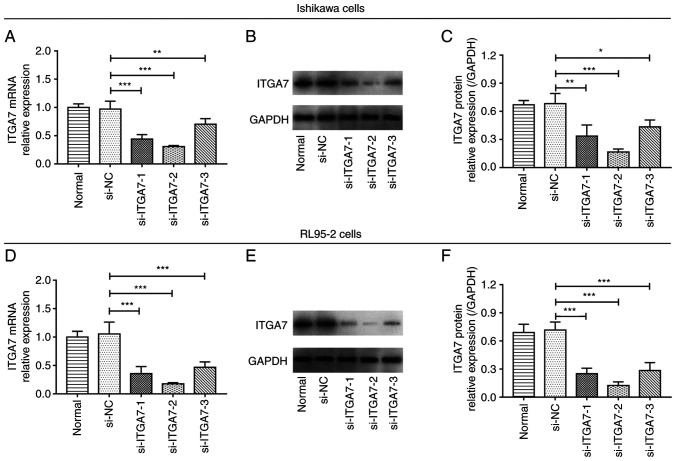
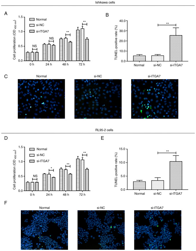
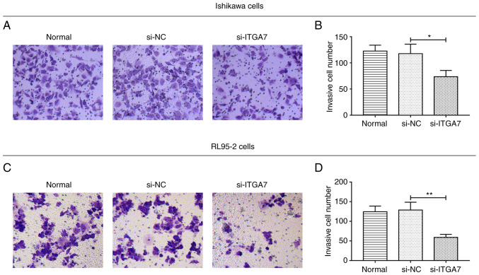
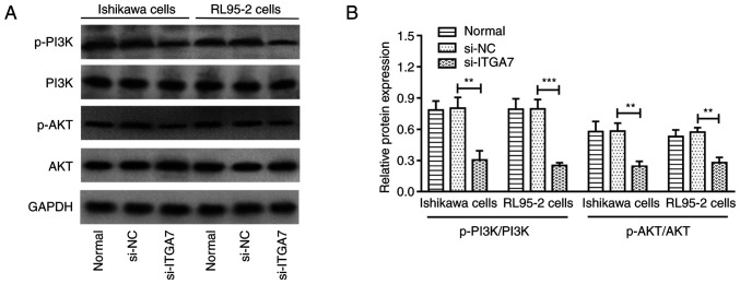
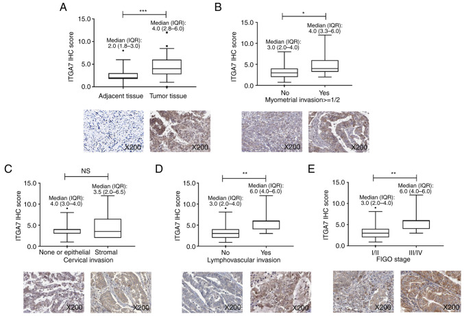
Targeting integrin α7 (ITGA7) suppresses malignant progression of several types of cancer, including tongue squamous cell carcinoma, hepatocellular carcinoma and non-small cell lung cancer, while the effect of its knockdown on cell function and its association with clinicopathological features in endometrial cancer (EC) is unclear. The present study aimed to investigate this issue. ITGA7 was knocked down by short-interfering (si)RNA in Ishikawa and RL95-2 cells followed by western blotting and reverse transcription-quantitative PCR assays. Subsequently, cell proliferation, apoptosis, invasion and expression levels of PI3K, phosphorylated (p-) PI3K, AKT and p-AKT were determined using Cell Counting Kit-8, TUNEL, Transwell assays and western blotting. Moreover, ITGA7 in tumor and adjacent tissues from 50 patients with endometrial cancer was detected using immunohistochemical assay. ITGA7 expression was increased in EC cell lines (HEC-1A, RL95-2, Ishikawa and KLE) compared with telomerase-immortalized human endometrial stromal cells (THESCs). In both Ishikawa and RL95-2 cells, three ITGA7 siRNAs all demonstrated good efficiency on ITGA7 knockdown, amongst which the one with the highest efficiency was selected for the following experiments. ITGA7 knockdown reduced cell proliferation and invasion, while inducing apoptosis; moreover, it suppressed p-PI3K/PI3K and p-AKT/AKT ratios. In patients with EC, ITGA7 expression was increased in tumor tissues compared with adjacent tissues, and its lower tumor expression was associated with myometrial invasion (<1/2), non-lymphovascular invasion and decreased FIGO stage. In conclusion, ITGA7 knockdown repressed proliferation, invasion and the PI3K/AKT pathway while inducing apoptosis in EC cell lines, and its insufficiency was associated with less advanced tumor features in EC patients. These results indicated that ITGA7 may be a potential target for the treatment of EC.
Keywords: PI3K/AKT pathway; cell function; clinicopathological features; endometrial cancer; integrin α7.
Copyright: © Liang et al.
Conflict of interest statement
The authors declare that they have no competing interests.
Figures
Similar articles
-
Integrin alpha 7 correlates with poor clinical outcomes, and it regulates cell proliferation, apoptosis and stemness via PTK2-PI3K-Akt signaling pathway in hepatocellular carcinoma.Cell Signal. 2020 Feb;66:109465. doi: 10.1016/j.cellsig.2019.109465. Epub 2019 Nov 5. Cell Signal. 2020. PMID: 31698037
-
Integrin α7 correlates with worse clinical features and prognosis, and its knockdown inhibits cell proliferation and stemness in tongue squamous cell carcinoma.Int J Oncol. 2020 Jan;56(1):69-84. doi: 10.3892/ijo.2019.4927. Epub 2019 Nov 29. Int J Oncol. 2020. PMID: 31789398 Free PMC article.
-
[Preliminary investigation of the expression and functions of insulin receptor isoforms in endometrial carcinoma].Zhonghua Fu Chan Ke Za Zhi. 2012 Nov;47(11):839-45. Zhonghua Fu Chan Ke Za Zhi. 2012. PMID: 23302125 Chinese.
-
Integrin α7 knockdown suppresses cell proliferation, migration, invasion and EMT in hepatocellular carcinoma.Exp Ther Med. 2021 Apr;21(4):309. doi: 10.3892/etm.2021.9740. Epub 2021 Feb 1. Exp Ther Med. 2021. PMID: 33717252 Free PMC article.
-
Integrin α7 high expression correlates with deteriorative tumor features and worse overall survival, and its knockdown inhibits cell proliferation and invasion but increases apoptosis in breast cancer.J Clin Lab Anal. 2019 Oct;33(8):e22979. doi: 10.1002/jcla.22979. Epub 2019 Jul 19. J Clin Lab Anal. 2019. PMID: 31325216 Free PMC article.
Cited by
-
Molecular typing of gliomas on the basis of integrin family genes and a functional study of ITGA7.Sci Rep. 2025 Apr 10;15(1):12306. doi: 10.1038/s41598-025-97342-3. Sci Rep. 2025. PMID: 40210748 Free PMC article.
-
Breast cancer-derived CAV1 promotes lung metastasis by regulating integrin α6β4 and the recruitment and polarization of tumor-associated neutrophils.Int J Biol Sci. 2024 Oct 14;20(14):5695-5714. doi: 10.7150/ijbs.94153. eCollection 2024. Int J Biol Sci. 2024. PMID: 39494337 Free PMC article.
-
ZNF714 Supports Pro-Oncogenic Features in Lung Cancer Cells.Int J Mol Sci. 2023 Oct 24;24(21):15530. doi: 10.3390/ijms242115530. Int J Mol Sci. 2023. PMID: 37958512 Free PMC article.
References
LinkOut - more resources
Full Text Sources