. 2022 Oct 25;4(1):vdac170.
doi: 10.1093/noajnl/vdac170. eCollection 2022 Jan-Dec.

Diagnostics and prospective outcome of a diffuse glioneuronal tumor with oligodendroglioma-like features and nuclear clusters after surgical resection (DGONC): a case report

Affiliations

Diagnostics and prospective outcome of a diffuse glioneuronal tumor with oligodendroglioma-like features and nuclear clusters after surgical resection (DGONC): a case report

Chelsea Howie et al. Neurooncol Adv. .
No abstract available

Keywords: brain tumor; molecular diagnostics; patient outcomes.


Figures

Figure 1.
Figure 1.
Radiologic features of tumor. (A-D) Axial T2, Coronal T1 post contrast, Axial ADC, and Axial SWAN images of the lesion at presentation. (A) Lesion is well defined, encasing the right MCA. (B) Demonstrates no enhancement post contrast administration. (C) No diffusion restriction noted. (D) Central calcifications seen on susceptibility weighted images. (E-H) Follow-up axial T2 images. (E) Subtotal resection of the tumor after first surgery. (F) Small residual component posterior to the MCA after second surgery. (G) Stable residual lesion on follow-up scans at 3 months, and 1-year post-surgery (H).
Figure 2.
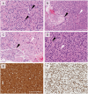
Figure 2.
Histopathologic features of tumor. (A) Moderately cellular tumor with round nuclei, perinuclear halos, and nuclear clusters (black arrows). (B) Areas of mucinous degeneration with foam cells (black arrow) and scattered microvascular proliferation (white arrow). (C) Areas of confluent apoptosis (black arrow) and associated endothelial proliferation (white arrow). (D) Frequent entrapped normal neurons (black arrow) and mitotic figures (white arrow). (E) Strong diffuse positivity for synaptophysin. (F) Strong diffuse nuclear staining for OLIG2. Scale bar represents 200 microns.
Figure 3.
Figure 3.
Molecular features of tumor. (A) Copy number variation plot showing the characteristic loss of chromosome 14. (B) tSNE embedding of the index case in regard to selected tumor types central neuroblastoma (CN), diffuse glioneuronal tumors with oligodendroglioma-like features and nuclear clusters (DGONC), Diffuse leptomeningeal glioneuronal tumor subgroup 1 and 2 (DLGNT_1 and _2), dysembryoplastic neuroepithelial tumor (DNET), extraventricular neurocytoma (EVNCYT), ganglioglioma (GG), desmoplastic infantile ganglioglioma/astrocytoma (LGG_DIG_DIA), oligodendroglioma (O_IDH), pilocytic astrocytoma cortical, infratentorial and midline (PA_Cort, _INF & _mid), rosette-forming glioneuronal tumor (RGNT).

References

    1. Louis DN, Perry A, Wesseling P, et al. . The 2021 WHO Classification of Tumors of the Central Nervous System: a summary. Neuro Oncol 2021;23(8):1231–1251. - PMC - PubMed
    1. Deng MY, Sill M, Sturm D, et al. . Diffuse glioneuronal tumour with oligodendroglioma-like features and nuclear clusters (DGONC) - a molecularly defined glioneuronal CNS tumour class displaying recurrent monosomy 14. Neuropathol Appl Neurobiol. 2020;46(5):422–430. - PubMed
    1. Pickles JC, Mankad K, Aizpurua M, et al. . A case series of diffuse glioneuronal tumours with oligodendroglioma-like features and nuclear clusters (DGONC). Neuropathol Appl Neurobiol. 2021;47(3):464–467. - PMC - PubMed
    1. Capper D, Jones DTW, Sill M, et al. . DNA methylation-based classification of central nervous system tumours. Nature 2018;555(7697):469–474. - PMC - PubMed
    1. Kamnasaran D, Cox DW. Current status of human chromosome 14. J Med Genet. 2002;39(2):81–90. - PMC - PubMed

LinkOut - more resources