. 2023 Feb 1;210(3):259-270.
doi: 10.4049/jimmunol.2200487.

Lung Resident Memory T Cells Activated by Oral Vaccination Afford Comprehensive Protection against Pneumonic Yersinia pestis Infection

Affiliations

Lung Resident Memory T Cells Activated by Oral Vaccination Afford Comprehensive Protection against Pneumonic Yersinia pestis Infection

Amit K Singh et al. J Immunol. .

Abstract

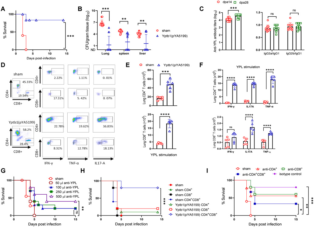
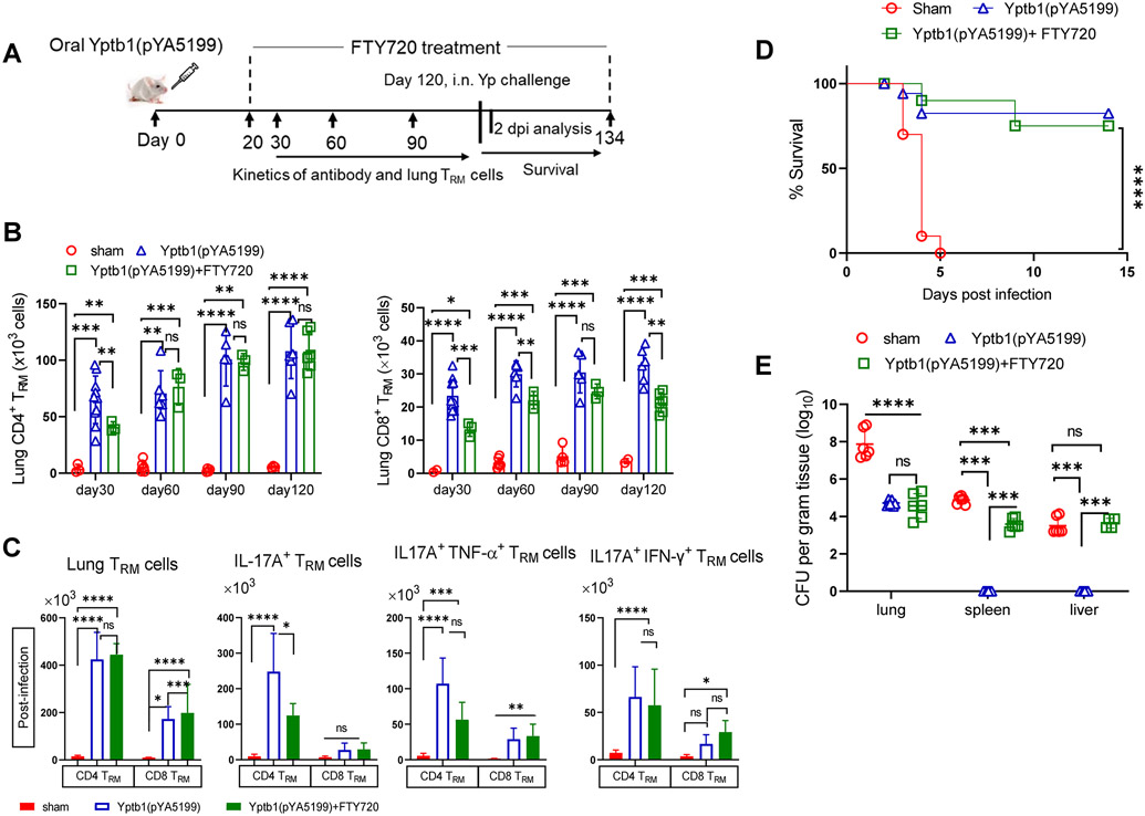
A growing body of evidence has shown that resident memory T (TRM) cells formed in tissue after mucosal infection or vaccination are crucial for counteracting reinfection by pathogens. However, whether lung TRM cells activated by oral immunization with Yptb1(pYA5199) play a protective role against pneumonic plague remains unclear. In this study, we demonstrated that lung CD4+ and CD8+ TRM cells significantly accumulated in the lungs of orally Yptb1(pYA5199)-vaccinated mice and dramatically expanded with elevated IL-17A, IFN-γ, and/or TNF-α production after pulmonary Yersinia pestis infection and afforded significant protection. Short-term or long-term treatment of immunized mice with FTY720 did not affect lung TRM cell formation and expansion or protection against pneumonic plague. Moreover, the intratracheal transfer of both lung CD4+ and CD8+ TRM cells conferred comprehensive protection against pneumonic plague in naive recipient mice. Lung TRM cell-mediated protection was dramatically abolished by the neutralization of both IFN-γ and IL-17A. Our findings reveal that lung TRM cells can be activated via oral Yptb1(pYA5199) vaccination, and that IL-17A and IFN-γ production play an essential role in adaptive immunity against pulmonary Y. pestis infection. This study highlights an important new target for developing an effective pneumonic plague vaccine.


Conflict of interest statement

Conflict of interest: All authors declare that they have no conflicts of interest.

Figures

Figure 1.
Figure 1.. Oral immunization with the Yptb1(pYA5199) vaccine confers T cell-mediated comprehensive protection against pneumonic Y. pestis infection.
(A) On the 42nd day after immunization, mice (n=15, mixed males and females) were intranasally challenged with 5 × 10³ CFU (50 LD50) of Y. pestis KIM6+(pCD1Ap). Survival was recorded for 14 dpi. (B) The bacterial burden in the lung, spleen, and liver at 2 dpi. (C) Antibody responses. Serum anti-YPL antibody titers in immunized mice at 14 and 28 dpv [left]; the ratios of IgG2a/IgG1 and IgG2b/IgG1 to YPL [right]. (D) Representative flow plots showing the frequency of lung CD4+ and CD8+ T cells and the respective cells producing IFN-γ, TNF-α, or IL-17A in mice (n=6 females). (E) At dpv 42, lung single cells obtained from Yptb1(pYA5199) and sham mice were in vitro stimulated for 72 h with YPL. The absolute number of lung CD4+ and CD8+ T cells and (F) lung T cells expressing IFN-γ, TNF-α, or IL-17A were determined by Flow cytometer. (G) Serum transfer. Naïve mice (n=10, females) were i.p. injected with different volumes of serum collected from sham and Yptb1(pYA5199)-immunized mice at 42 dpv. Twenty-four hours post-injection, recipient mice were intranasally challenged with 10 LD50 of Y. pestis. (H) Lung T-cell transfer. Naïve mice treated with a single dose of irradiation (5 Gy) were intravenously administered CD4+ and/or CD8+ T cells (2 × 10⁶ cells/mouse) isolated from sham or Yptb1(pYA5199)-immunized mice at 42 dpv. At 24 h post-administration, mice (n=10, females) were intranasally infected with 10 LD50 of Y. pestis, and survival was monitored for 14 days. (I) T-cell depletion. Mice (n=10, females) were depleted of CD4+ and/or CD8+ T cells by i.p. administration of anti-CD4, anti-CD8, or anti-CD4 plus anti-CD8 monoclonal antibodies (500 μg/each mouse in 200 μl) and then intranasally challenged with 50 LD50 of Y. pestis. Each symbol in the individual bar graph represents a data point obtained from an individual mouse. Each experiment was performed two times with identical conditions. Data obtained from experiments were pooled and analyzed and are presented as the mean ± SD. The statistical analysis is described in the Materials and Methods.
Figure 2.
Figure 2.. Lung CD4+ and CD8+ TRM cells are activated by oral Yptb1(pYA5199) immunization and expand after pulmonary Y. pestis infection.
Mice were intravenously injected via the tail vein with 5 μg of FITC-conjugated anti-CD45.2 mAbs diluted in 200 μl of sterile PBS to distinguish lung TRM cells from circulating T cells. Five minutes post-injection, mice were euthanized to isolate lung single cells (n=5 females). Lung cells isolated from Yptb1(pYA5199)-immunized and sham mice were in vitro stimulation with PMA+ ionomycin. (A) Representative flow plots of lung CD4+ and CD8+ TRM cells producing IFN-γ, TNF-α, and/or IL-17A in immunized and sham mice at 42 dpv. Lung TRM cells were gated based on CD45 (i.v. injection), CD4+/CD8+, CD44+, and CD69+, as well as intracellular cytokines (IFN-γ+, TNF-α+, and IL-17A+), in the flow cytometry protocol [top]. Quantitative analysis of the number of lung CD4+ or CD8+ TRM cells and corresponding cells producing IFN-γ, TNF-α, and/or IL-17A [bottom]. (B) Representative flow plots of lung CD4+ and CD8+ TRM cells in mice at 2 dpi [top]. Quantitative analysis of the number of lung CD4+ or CD8+ TRM cells and corresponding cells producing IFN-γ, TNF-α, and/or IL-17A (bottom) (n=10 females). (C) Kinetic analysis of lung CD4+ and CD8+ TRM cells after pulmonary Y. pestis infection (n=10 females). Data obtained from a minimum of two experiments are presented as the mean ± SD. The statistical analysis is described in the Materials and Methods.
Figure 3.
Figure 3.. CD4+ and CD8+ TRM cells established in the lungs of FTY720-treated Yptb1(pYA5199)-immunized mice offer protection against pulmonary Y. pestis infection.
(A) Scheme of the immunization, FTY720 treatment, TRM cell analysis, and Y. pestis infection. The lung single cell isolated Yptb1(pYA5199)+FTY720 immunized and sham mice were in vitro stimulated with PMA+ionomycine. (B) Quantitative analysis of the number of lung CD4+ or CD8+ TRM cells and corresponding cells producing IFN-γ, TNF-α, and/or IL-17A in FTY720-treated Yptb1(pYA5199)-immunized mice at 42 dpv (n=5 females). (C) Quantitative analysis of the number of lung CD4+ or CD8+ TRM cells and corresponding cells producing IFN-γ, TNF-α, and/or IL-17A in FTY720-treated Yptb1(pYA5199)-immunized mice at 2 dpi. (D) On 42 dpv, FTY720-treated Yptb1(pYA5199)-immunized mice (n=15, mixed males and females) were intranasally challenged with 50 LD50 of Y. pestis. Survival was recorded for 14 days. (E) The bacterial burden was evaluated in the lung, spleen, and liver at 2 dpi (n=6, mixed males and females). (F) Scheme of the FTY720 treatment 10 days before oral Yptb1(pYA5199) immunization and until the end of the experiment. (G) Quantitative analysis of the number of lung CD4+ or CD8+ TRM cells in pre- and post-FTY720-treated Yptb1(pYA5199)-immunized mice at 42 dpv. (H) On 42 dpv, mice (n=5, mixed males and females) were intranasally challenged with 50 LD50 of Y. pestis. (I) Adoptive transfer of FACS-sorted lung circulating (CD45+) T cells or TRM cells (CD45) from Yptb1(pYA5199)-immunized mice. Sorted cells were injected into irradiated (5 Gy) naïve mice via the intratracheal route. At 24 h after administration, recipient mice (n=10 females) were i.n. challenged with 10 LD50 of Y. pestis. In the bar plots, each symbol represents a data point obtained from an individual mouse. Data are presented as the mean ± SD. The statistical analysis is described in the Materials and Methods.
Figure 4.
Figure 4.. Local depletion of lung resident CD4+ and/or CD8+ T cells in Yptb1(pYA5199)-immunized mice impairs protection against pneumonic plague.
(A) Scheme of immunization, FTY720 treatment, T-cell depletion, and survival against pneumonic plague infection. (B) Survival study in the lung T cell-depleted mice. The Yptb1(pYA5199)+FTY720-immunized mice (n=6 females) were depleted of CD4+ and/or CD8+ T cells at the lung mucosal site by i.n. administration of 200 μg of anti-CD4, anti-CD8, or both mAbs and then intranasally challenged with 50 LD50 of Y. pestis. (C) Lung Y. pestis burden at 2 dpi (n= 5 females). (D) Representative flow plots showing the percentages of neutrophils and alveolar macrophages in the BAL fluid at 2 dpi. (E) The number of neutrophils and (F) alveolar macrophages (AMs) in the BAL fluid of control and T cell-depleted mice (n= 6 females) at 2 dpi. (G) Analysis of cytokines and chemokines in the BAL fluid samples collected at 2 dpi. Each symbol represents a data point obtained from an individual mouse. The statistical analysis is described in the Materials and Methods.
Figure 5.
Figure 5.. IFN-γ and IL-17A play a critical role in protection against pulmonary Y. pestis challenge.
(A) Scheme of neutralization of IFN-γ, IL-17A, or both in Yptb1(pYA5199)-immunized mice. Mice administrated with PBS (sham) were used as control. (B) Survival study with cytokine neutralization. The Yptb1(pYA5199)-immunized mice (n=10 females) were intraperitoneally injected with 200 μg of anti-IFN-γ, anti-IL-17A, or both mAbs and then intranasally challenged with 50 LD50 of Y. pestis. (C) Lung Y. pestis burden in the respective group of mice at 2 dpi. (D) The number of neutrophils and (E) AMs in the BAL fluid. (F) The absolute number of lung neutrophil and (G) AMs in the lung of mice (n=6 females) treated with anti-IFN-γ, anti-IL-17A, anti-IFN-γ/IL-17A, or isotype control antibodies at 2 dpi. (H) The number of CD4+ and (I) CD8+ TRM cells in the lungs of mice (n=6 females) treated with the respective antibodies at 2 dpi. (J) Analysis of cytokines and chemokines in the BAL fluid of mice (n=6 females) treated with the respective antibodies at 2 dpi. (K) Representative H&E-stained lung sections of oral Yptb1(pYA5199)-immunized mice treated with anti-IFN-γ, anti-IL-17A, anti-IFN-γ/IL-17A or isotype control antibodies, or sham mice collected at 2 dpi. The black arrow indicates a reduced alveolar lacunar space, while the red arrow indicates a lung lesion or hemorrhage. Each symbol represents a data point obtained from an individual mouse. Data are presented as the mean ± SD. The statistical analysis is described in the Materials and Methods.
Figure 6.
Figure 6.. Lung TRM cells activated by oral Yptb1(pYA5199) immunization confer long-lasting protection.
(A) Scheme of the immunization, immune parameter analysis, and challenge in the long-term animal study. (B) Kinetic analysis of lung CD4+ and CD8+ TRM cells in Yptb1(pYA5199)-immunized mice treated with or without FTY720 at 30, 60, 90, and 120 dpv. (n=6 females) (C) Mice were intravenously injected via the tail vein with 5 μg of FITC-conjugated anti-CD45.2 mAbs diluted in 200 μl of sterile PBS to distinguish lung TRM cells from circulating T cells. Five minutes post-injection, mice were euthanized to isolate lung single cells (n=5 females). Lung cells isolated from Yptb1(pYA5199), Yptb1(pYA5199)+FTY720 immunized and sham mice were in vitro stimulation with PMA+ ionomycin. Quantitative analysis of the number of lung CD4+ or CD8+ TRM cells and corresponding cells producing IFN-γ, TNF-α, and/or IL-17A in Yptb1(pYA5199)-immunized mice treated with or without FTY720 and control mice was performed at 2 dpi. (D) On the 120th day after immunization, mice (n=10, equal number of males and females) were intranasally challenged with 50 LD50 of Y. pestis. (E) The bacterial burden was evaluated in the lung, liver, and spleen of mice (n=6, mixed males and females) at 2 dpi. Data obtained from experiments are presented as the mean ± SD. The statistical analysis is described in Materials and Methods.

Similar articles

Cited by

References

    1. Pechous RD, Sivaraman V, Stasulli NM, and Goldman WE. 2016. Pneumonic Plague: The darker side of Yersinia pestis. Trends Microbiol 24: 190–197. - PubMed
    1. Alderson J, Quastel M, Wilson E, and Bellamy D. 2020. Factors influencing the re-emergence of plague in Madagascar. Emerg Top Life Sci 4: 411–421. - PMC - PubMed
    1. Inglesby TV, Dennis DT, Henderson DA, Bartlett JG, Ascher MS, Eitzen E, Fine AD, Friedlander AM, Hauer J, Koerner JF, Layton M, McDade J, Osterholm MT, O'Toole T, Parker G, Perl TM, Russell PK, Schoch-Spana M, Tonat K, and Biodefense WGC. 2000. Plague as a biological weapon - Medical and public health management. Jama-J Am Med Assoc 283: 2281–2290. - PubMed
    1. Sun W, and Singh AK. 2019. Plague vaccine: recent progress and prospects. NPJ Vaccines 4: 11. - PMC - PubMed
    1. 2018. Efficacy trials of Plague Vaccines: endpoints, trial design, site selection. In WHO Workshop 2018. Efficacy trials of Plague Vaccines: endpoints, trial design, site selection, INSERM, Paris, France.

Publication types