

Electrocardiographic imaging demonstrates electrical synchrony improvement by dynamic atrioventricular delays in patients with left bundle branch block and preserved atrioventricular conduction
- PMID: 36480445
- PMCID: PMC9935053
- DOI: 10.1093/europace/euac224
Electrocardiographic imaging demonstrates electrical synchrony improvement by dynamic atrioventricular delays in patients with left bundle branch block and preserved atrioventricular conduction
Abstract
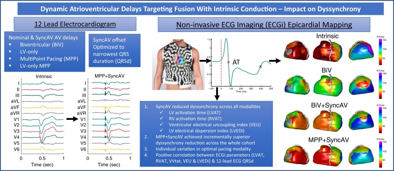
Aims: Cardiac resynchronization therapy programmed to dynamically fuse pacing with intrinsic conduction using atrioventricular (AV) timing algorithms (e.g. SyncAV) has shown promise; however, mechanistic data are lacking. This study assessed the impact of SyncAV on electrical dyssynchrony across various pacing modalities using non-invasive epicardial electrocardiographic imaging (ECGi).
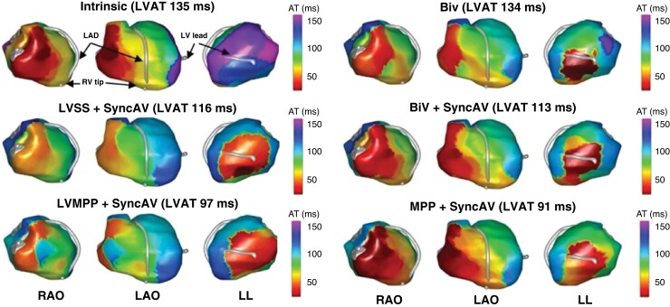
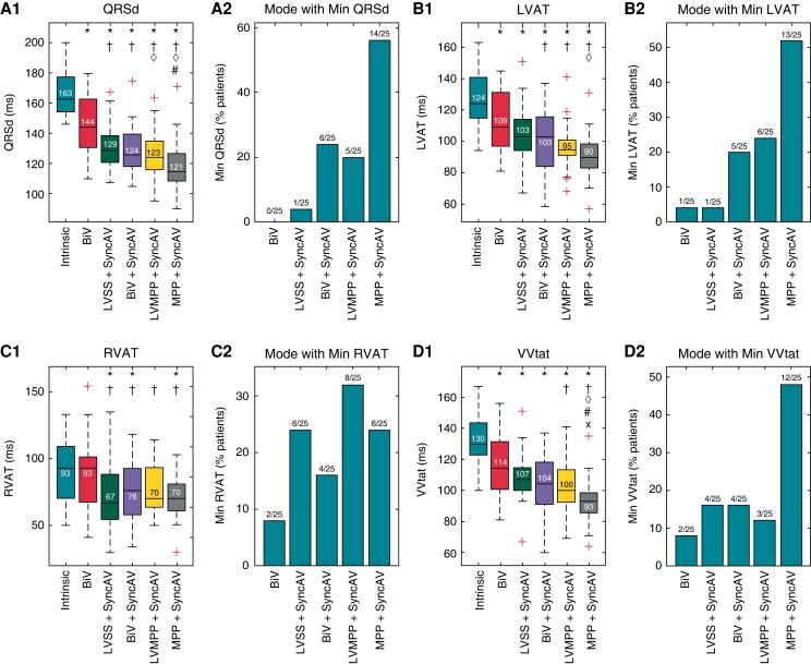
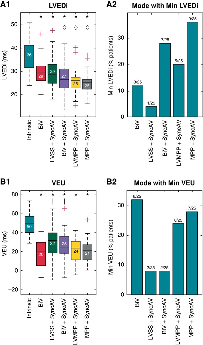
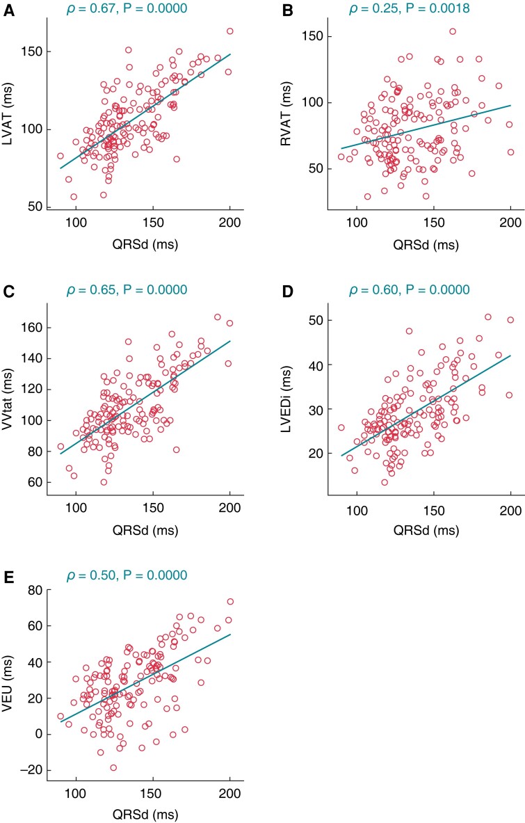
Methods and results: Twenty-five patients with left bundle-branch block (median QRS duration (QRSd) 162.7 ms) and intact AV conduction (PR interval 174.0 ms) were prospectively enrolled. ECGi was performed acutely during biventricular pacing with fixed nominal AV delays (BiV) and using SyncAV (optimized for the narrowest QRSd) during: BiV + SyncAV, LV-only single-site (LVSS + SyncAV), MultiPoint pacing (MPP + SyncAV), and LV-only MPP (LVMPP + SyncAV). Dyssynchrony was quantified via ECGi (LV activation time, LVAT; RV activation time, RVAT; LV electrical dispersion index, LVEDi; ventricular electrical uncoupling index, VEU; and biventricular total activation time, VVtat). Intrinsic conduction LVAT (124 ms) was significantly reduced by BiV pacing (109 ms) (P = 0.001) and further reduced by LVSS + SyncAV (103 ms), BiV + SyncAV (103 ms), LVMPP + SyncAV (95 ms), and MPP + SyncAV (90 ms). Intrinsic RVAT (93 ms), VVtat (130 ms), LVEDi (36 ms), VEU (50 ms), and QRSd (163 ms) were reduced by SyncAV across all pacing modes. More patients exhibited minimal LVAT, VVtat, LVEDi, and QRSd with MPP + SyncAV than any other modality.
Conclusion: Dynamic AV delay programming targeting fusion with intrinsic conduction significantly reduced dyssynchrony, as quantified by ECGi and QRSd for all evaluated pacing modes. MPP + SyncAV achieved the greatest synchrony overall but not for all patients, highlighting the value of pacing mode individualization during fusion optimization.
Keywords: Atrioventricular delay; CRT optimization; Electrocardiographic imaging; Fusion pacing; MultiPoint pacing; SyncAV.
© The Author(s) 2022. Published by Oxford University Press on behalf of the European Society of Cardiology.
Conflict of interest statement
Conflict of interest: N.B., L.C.M., and J.O.M. are employees of Abbott.
Figures
References
-
- Cleland JG, Abraham WT, Linde C, Gold MR, Young JB, Claude Daubert J, et al. An individual patient meta-analysis of five randomized trials assessing the effects of cardiac resynchronization therapy on morbidity and mortality in patients with symptomatic heart failure. Eur Heart J 2013;34:3547–56. - PMC - PubMed
-
- O'Donnell D, Wisnoskey B, Badie N, Odgers L, Smart T, Ord M, et al. Electrical synchronization achieved by multipoint pacing combined with dynamic atrioventricular delay. J Interv Card Electrophysiol 2021;61:453–460. - PubMed
-
- Thibault B, Ritter P, Bode K, Calo L, Mondesert B, Mangual JO, et al. Dynamic programming of atrioventricular delay improves electrical synchrony in a multicenter cardiac resynchronization therapy study. Heart Rhythm 2019;16:1047–56. - PubMed
MeSH terms
Grants and funding
LinkOut - more resources
Full Text Sources
Medical
Research Materials
