. 2022 Dec 13;119(50):e2210669119.
doi: 10.1073/pnas.2210669119. Epub 2022 Dec 8.

Biophysical characterization of calcium-binding and modulatory-domain dynamics in a pentameric ligand-gated ion channel

Affiliations

Biophysical characterization of calcium-binding and modulatory-domain dynamics in a pentameric ligand-gated ion channel

Marie Lycksell et al. Proc Natl Acad Sci U S A. .

Abstract

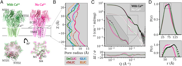
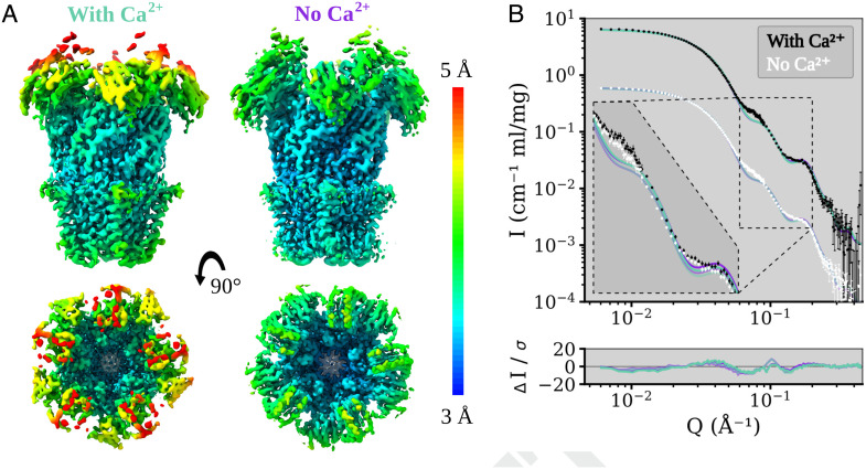
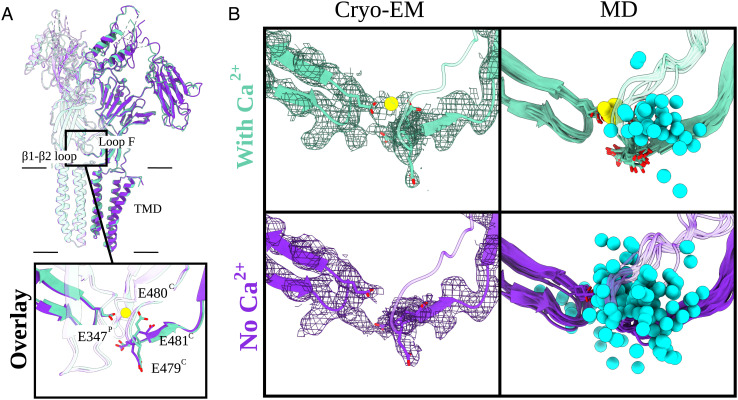
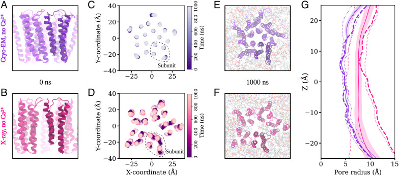
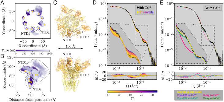
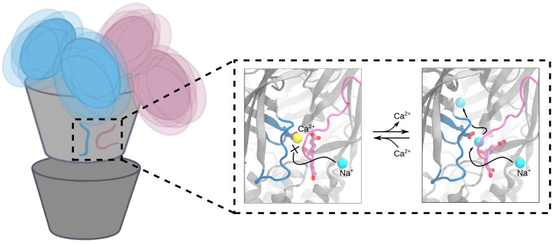
Pentameric ligand-gated ion channels (pLGICs) perform electrochemical signal transduction in organisms ranging from bacteria to humans. Among the prokaryotic pLGICs, there is architectural diversity involving N-terminal domains (NTDs) not found in eukaryotic relatives, exemplified by the calcium-sensitive channel (DeCLIC) from a Desulfofustis deltaproteobacterium, which has an NTD in addition to the canonical pLGIC structure. Here, we have characterized the structure and dynamics of DeCLIC through cryoelectron microscopy (cryo-EM), small-angle neutron scattering (SANS), and molecular dynamics (MD) simulations. In the presence and absence of calcium, cryo-EM yielded structures with alternative conformations of the calcium-binding site. SANS profiles further revealed conformational diversity at room temperature beyond that observed in static structures, shown through MD to be largely attributable to rigid-body motions of the NTD relative to the protein core, with expanded and asymmetric conformations improving the fit of the SANS data. This work reveals the range of motion available to the DeCLIC NTD and calcium-binding site, expanding the conformational landscape of the pLGIC family. Further, these findings demonstrate the power of combining low-resolution scattering, high-resolution structural, and MD simulation data to elucidate interfacial interactions that are highly conserved in the pLGIC family.

Keywords: Cys-loop receptors; calcium; ligand-gated ion channel; small-angle neutron scattering.


Conflict of interest statement

The authors declare no competing interest.

Figures

Fig. 1.
Fig. 1.
Structure overview of DeCLIC and the fit of the DeCLIC X-ray structures to small-angle neutron scattering data. (A) The structure of DeCLIC consists of two beta-rich N-terminal domains (NTD1 and NTD2), a beta-sandwich extracellular domain (ECD) with a calcium-binding site, and a transmembrane domain (TMD) with four helices M1 through M4. X-ray crystallography structures have been determined by Hu et al. (11) in the presence (green) and absence (pink) of calcium, with the absence of calcium leading to a conformation with a more contracted NTD and a more expanded TMD. (B) Pore profiles of the TMD region in DeCLIC (green and pink, solid lines) and of the related channels, GLIC (dark and light blue, dashed lines), sTeLIC (dark red, dash-dot line), and ELIC (orange, dotted line). In the presence of calcium, DeCLIC (green line) has a contracted pore similar to ELIC (orange dots) and GLIC at resting conditions (dark blue dashes). In the absence of calcium, DeCLIC (pink line) has a wide pore at the 9’ hydrophobic gate (Z = 0 Å), similar to sTeLIC (dark red dash-dots), and is at the 16’ contraction (Z  ≈  10 Å) similar to GLIC at activating conditions (light blue dashes). (C) Small-angle neutron scattering from DeCLIC in the presence (black) and absence (white) of calcium, with predicted curves from the crystal structures in the presence (green) and absence (pink) of calcium. The dataset in the presence of calcium has been offset by a factor of 10 for clarity. The inset shows the data on the same scale zoomed in on Q ∈ [0.06, 0.2] Å−1, with darker curves fitted to the SANS data with calcium and paler curves fitted to the calcium-free SANS data. The calcium-free crystal structure has poor goodness of fit (χ2 > 60) to the SANS data, while the calcium-containing crystal structure gives moderate goodness of fit (χ2 of 10.8 to SANS with calcium and χ2 of 8.8 to SANS without calcium), deviating from the experimental data mainly for Q ∈ [0.06, 0.09] Å−1. (D) Pair distance distribution of DeCLIC from SANS with (black) and without (white) calcium and from crystal structures with (green) and without (pink) calcium. There is close agreement between the distribution from SANS with calcium and from the calcium-containing crystal structure, while the SANS data collected in the absence of calcium have features similar to those of both crystal structures (zoom box).
Fig. 2.
Fig. 2.
Local resolution coloring of the DeCLIC Cryo-EM reconstructions in the presence and absence of calcium and their agreement with the SANS data. (A) Cryo-EM densities of DeCLIC in the presence of 10 mM Ca2+ (Left), and from a Ca2+ chelated condition (Right). The densities were resolved to over all resolutions of 3.5 Å (condition with Ca2+) and 3.2 Å (condition with no Ca2+) respectively. Density is colored by local resolution according to the scale bar to the right. The quality is worse in the NTD region of the protein compared to that of the TMD or ECD. (B) Fits of model spectra calculated from the two cryo-EM structures in the presence (aquamarine) and absence (purple) of Ca2+ to SANS data collected with Ca2+ (black) and with no Ca2+ (white). The structures yield similar goodness of fit, χ2∈[11.2, 12.1]. SANS data with Ca2+ are offset by a factor of 10 for easier visualization. The inset shows the data on the same scale zoomed on Q ∈ [0.06, 0.2] Å−1, with darker curves fitted to the with-calcium SANS data and paler curves fitted to the calcium-free SANS data.
Fig. 3.
Fig. 3.
Arrangement of the calcium-binding residues elucidated by cryo-EM and MD. (A) Overlay of two adjacent subunits of DeCLIC cryo-EM structures with and without Ca2+ (aquamarine and purple, respectively). Protein domains and main Ca2+-binding motifs are shown. Those include the transmembrane domain (TMD), extracellular domain (ECD), and two amino-terminal domains (NTD1 and NTD2) as well as the β1 to β2 loop from the principal subunit (P) and loop F from the complementary subunit (C). The boxed-in view (Bottom) shows the relevant side chains (sticks, colored by heteroatom). The Ca2+ ion present in the Ca2+ dataset (aquamarine) is colored in yellow. (B) Cryo-EM panels (Left) represent the cryo-EM structures and their corresponding densities (mesh at α  =  0.012 to 0.015). Relevant side chains around the Ca2+-binding site [loop F (C), β1 to β2 loop (P)] are displayed as sticks (heteroatom coloring). The density for the Ca2+-binding residues as well as for the Ca2+ ion (in the with-Ca2+ condition) is well resolved. MD panels (Right) illustrate eleven individual conformations (one snapshot for every 100 ns) from 1-μs long MD simulations of the cryo-EM models. The relevant Ca2+ residues are displayed as sticks (colored by heteroatom). In cyan, we have Na+ ions (sphere, trajectories generated for every 10 ns) and Ca2+ ion in yellow. In the no-Ca2+ condition, the Na+ ions are free to enter the cavity which is occupied by Ca2+ in the with-Ca2+ condition.
Fig. 4.
Fig. 4.
Behavior of the transmembrane domain in molecular dynamics simulations. (A and B) View of the transmembrane region of DeCLIC, seen from the membrane plane, prior to MD simulation in the calcium-free cryo-EM structure and in the calcium-free X-ray structure. (C and D) Position of the center of mass of each transmembrane helix in the bilayer plane through 1,000 ns of MD simulations and four replicas. For the calcium-free cryo-EM structure, the helices of the transmembrane region remain close to their positions in the structure, while the transmembrane domain in simulation of the X-ray structure without calcium undergoes a conformational change to an asymmetric arrangement of the subunits. (E and F) Transmembrane region seen from the extracellular side at the end of the simulations. The cryo-EM structure without calcium has retained a symmetric arrangement, while the X-ray structure without calcium has become asymmetric, with lipids penetrating to the pore in one simulation replica. (G) Pore profiles tracing the protein backbone for the calcium-free cryo-EM (purple) and X-ray (pink) systems. Dashed lines show the profile for the starting structure, solid lines the simulation average with SD in colored fill, and dotted lines minimum and maximal values during the simulations. There is little variation in the pore profile of the cryo-EM system during the simulations, while the wide lower part of the pore in the calcium-free X-ray structure contracts during the simulations.
Fig. 5.
Fig. 5.
The N-terminal domains of DeCLIC are highly dynamic, and an asymmetric arrangement thereof with a mix of contracted and extended NTD positions yields the best fit to the SANS data. (A and B) Position of the center of mass of each NTD lobe over time in four MD simulations, showing their positions in the XY-plane (A) and their Z-coordinate as a function of distance from the pore axis (B). NTD1 is highly mobile, sampling positions further out, lower down, and higher up than the starting conformation. NTD2 has less mobility, mainly sampling positions along Z. Yellow dots show the position of the NTD lobes in C. (C) Snapshot of the MD simulation frame yielding the best fit to the with-calcium SANS data (χ2 of 5.2), seen from the extracellular side (Top) and from the plane of the membrane (Bottom). The protein has adopted an asymmetric conformation with three NTD1 domains in positions similar to the determined structures and two NTD1 lobes further out and down. The snapshot is from the simulations started from the no-Ca2+ cryo-EM structure. (D) Fits of snapshots from simulations of the no-Ca2+ cryo-EM structure and the error-weighted residual between the models and the scattering profile. The simulations yield theoretical distributions around the experimental scattering curve, containing models with better fit (best χ2 of 5.2) to the data than the experimental structure from which the simulations were launched (χ2 of 11.2). Inset shows Q ∈ [0.06, 0.2] Å−1. (E) Comparison between the best-fitting model from simulations (yellow), structures of closed-like DeCLIC (purple, aquamarine, and green; note that curves overlap), and the X-ray no-Ca2+ structure (pink). As seen in the inset showing Q ∈ [0.06, 0.2] Å−1, the simulation model has the best fit to both features in the scattering profile.
Fig. 6.
Fig. 6.
Conformational variability and proposed calcium-binding site behavior in closed DeCLIC. The positions of the N-terminal domains of DeCLIC fluctuate around the core of the protein, sampling a wide range of conformations where the average can be described by an asymmetric conformation with a mix of compact and extended NTD positions. In the calcium-binding site, bound calcium blocks sodium access to the site. Following calcium depletion, sodium can enter the central pore through the calcium-binding site. The calcium-binding site is thus proposed to act as a supplementary way for sodium ions to reach the ion conduction pathway, with the three consecutive glutamates on loop F shepherding sodium from outside the protein, through the calcium-binding site, and into the extracellular part of the ion channel pore.
Fig. 7.
Fig. 7.
Charged residues at the subunit interface present in other family members. (A) Overlay of the subunit interface (loop F, Cys/Pro-loop, and β1 to β2 loop) from DeCLIC with other family members, nAChR (sky blue), ELIC (gray), 5-HT3AR (pink), and GABAAR (salmon) with Ca2+ ion in yellow and Ba2+ in green. The important charged residues are displayed as sticks (colored by heteroatom). (B) Display of each family member from (A) individually for clearer representation with relevant charged residues displayed as sticks and colored by heteroatom.

References

    1. Tasneem A., Iyer L. M., Jakobsson E., Aravind L., Identification of the prokaryotic ligand-gated ion channels and their implications for the mechanisms and origins of animal Cys-loop ion channels. Genome Biol. 6, 1–12 (2005). - PMC - PubMed
    1. Nemecz Á., Prevost M. S., Menny A., Corringer P. J., Emerging molecular mechanisms of signal transduction in pentameric ligand-gated ion channels. Neuron 90, 452–470 (2016). - PubMed
    1. Sauguet L., Shahsavar A., Delarue M., Crystallographic studies of pharmacological sites in pentameric ligand-gated ion channels. Biochim. Biophysi. Acta (BBA)-Gen. Subj. 1850, 511–523 (2015). - PubMed
    1. Taly A., Corringer P. J., Guedin D., Lestage P., Changeux J. P., Nicotinic receptors: Allosteric transitions and therapeutic targets in the nervous system. Nat. Rev. Drug Dis. 8, 733–750 (2009). - PubMed
    1. Kim J. J., et al. , Shared structural mechanisms of general anaesthetics and benzodiazepines. Nature 585, 303–308 (2020). - PMC - PubMed

Publication types

LinkOut - more resources