

Triggering of lymphocytes by CD28, 4-1BB, and PD-1 checkpoints to enhance the immune response capacities
- PMID: 36480493
- PMCID: PMC9731445
- DOI: 10.1371/journal.pone.0275777
Triggering of lymphocytes by CD28, 4-1BB, and PD-1 checkpoints to enhance the immune response capacities
Abstract
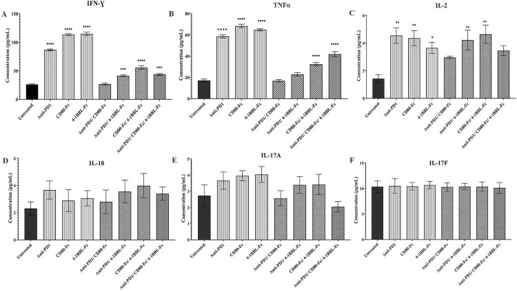
Tumor infiltrating lymphocytes (TILs) usually become exhausted and dysfunctional owing to chronic contact with tumor cells and overexpression of multiple inhibitor receptors. Activation of TILs by targeting the inhibitory and stimulatory checkpoints has emerged as one of the most promising immunotherapy prospectively. We investigated whether triggering of CD28, 4-1BB, and PD-1 checkpoints simultaneously or alone could enhance the immune response capacity of lymphocytes. In this regard, anti-PD-1, CD80-Fc, and 4-1BBL-Fc proteins were designed and produced in CHO-K1 cells as an expression host. Following confirmation of the Fc fusion proteins' ability to bind to native targets expressed on engineered CHO-K1 cells (CHO-K1/hPD-1, CHO-K1/hCD28, CHO-K1/hCTLA4, and CHO-K1/h4-1BB), the effects of each protein, on its own and in various combinations, were assessed in vitro on T cell proliferation, cytotoxicity, and cytokines secretion using the Mixed lymphocyte reaction (MLR) assay, 7-AAD/CFSE cell-mediated cytotoxicity assay, and a LEGENDplex™ Human Th Cytokine Panel, respectively. MLR results demonstrated that T cell proliferation in the presence of the combinations of anti-PD-1/CD80-Fc, CD80-Fc/4-1BBL-Fc, and anti-PD-1/CD80-Fc/4-1BBL-Fc proteins was significantly higher than in the untreated condition (1.83-, 1.91-, and 2.02-fold respectively). Furthermore, anti-PD-1 (17%), 4-1BBL-Fc (19.2%), anti-PD-1/CD80-Fc (18.6%), anti-PD-1/4-1BBL-Fc (21%), CD80-Fc/4-1BBL-Fc (18.5%), and anti-PD-1/CD80-Fc/4-1BBL-Fc (17.3%) significantly enhanced cytotoxicity activity compared to untreated condition (7.8%). However, concerning the cytokine production, CD80-Fc and 4-1BBL-Fc alone or in combination significantly increased the secretion of IFN-γ, TNF-α, and IL-2 compared with the untreated conditions. In conclusion, this research establishes that the various combinations of produced anti-PD-1, CD80-Fc, and 4-1BBL-Fc proteins can noticeably induce the immune response in vitro. Each of these combinations may be effective in killing or destroying cancer cells depending on the type and stage of cancer.
Copyright: © 2022 Kaviani et al. This is an open access article distributed under the terms of the Creative Commons Attribution License, which permits unrestricted use, distribution, and reproduction in any medium, provided the original author and source are credited.
Conflict of interest statement
The authors have declared that no competing interests exist.
Figures


Similar articles
-
Co-stimulation with 4-1BB ligand allows extended T-cell proliferation, synergizes with CD80/CD86 and can reactivate anergic T cells.Int Immunol. 2007 Dec;19(12):1383-94. doi: 10.1093/intimm/dxm106. Epub 2007 Oct 31. Int Immunol. 2007. PMID: 17977894
-
Changes in lymphocytes' telomerase activity by 4-1BB costimulation.J Cancer Res Ther. 2014 Oct-Dec;10(4):998-1003. doi: 10.4103/0973-1482.137906. J Cancer Res Ther. 2014. PMID: 25579543
-
4-1BB ligand induces cell division, sustains survival, and enhances effector function of CD4 and CD8 T cells with similar efficacy.J Immunol. 2001 Aug 1;167(3):1313-24. doi: 10.4049/jimmunol.167.3.1313. J Immunol. 2001. PMID: 11466348
-
CD80-Fc fusion protein as a potential cancer immunotherapy strategy.Antib Ther. 2023 Nov 30;7(1):28-36. doi: 10.1093/abt/tbad029. eCollection 2024 Jan. Antib Ther. 2023. PMID: 38235375 Free PMC article. Review.
-
Novel strategies for inhibiting PD-1 pathway-mediated immune suppression while simultaneously delivering activating signals to tumor-reactive T cells.Cancer Immunol Immunother. 2015 Oct;64(10):1287-93. doi: 10.1007/s00262-015-1677-5. Epub 2015 Mar 20. Cancer Immunol Immunother. 2015. PMID: 25792524 Free PMC article. Review.
Cited by
-
Parkinson's disease: exploring the systemic immune mechanisms through molecular investigations.Inflammopharmacology. 2025 Jul;33(7):3679-3699. doi: 10.1007/s10787-025-01816-9. Epub 2025 Jun 23. Inflammopharmacology. 2025. PMID: 40549315 Review.
-
Regulating the regulatory T cells as cell therapies in autoimmunity and cancer.Front Med (Lausanne). 2023 Sep 27;10:1244298. doi: 10.3389/fmed.2023.1244298. eCollection 2023. Front Med (Lausanne). 2023. PMID: 37828948 Free PMC article. Review.
-
The roles and molecular mechanisms of non-coding RNA in cancer metabolic reprogramming.Cancer Cell Int. 2024 Jan 18;24(1):37. doi: 10.1186/s12935-023-03186-0. Cancer Cell Int. 2024. PMID: 38238756 Free PMC article. Review.
-
Identification of Novel Molecular Panel as Potential Biomarkers of PAN-Gastrointestinal Cancer Screening: Bioinformatics and Experimental Analysis.Biology (Basel). 2025 Jul 2;14(7):803. doi: 10.3390/biology14070803. Biology (Basel). 2025. PMID: 40723362 Free PMC article.
References
Publication types
MeSH terms
Substances
LinkOut - more resources
Full Text Sources
Research Materials
