. 2022 Dec 8;17(12):e0277492.
doi: 10.1371/journal.pone.0277492. eCollection 2022.

Disseminated Intravascular Coagulation (DIC): Old player creates new perspectives on the polymicrobial sepsis model of CASP

Affiliations

Disseminated Intravascular Coagulation (DIC): Old player creates new perspectives on the polymicrobial sepsis model of CASP

Julia van der Linde et al. PLoS One. .

Abstract

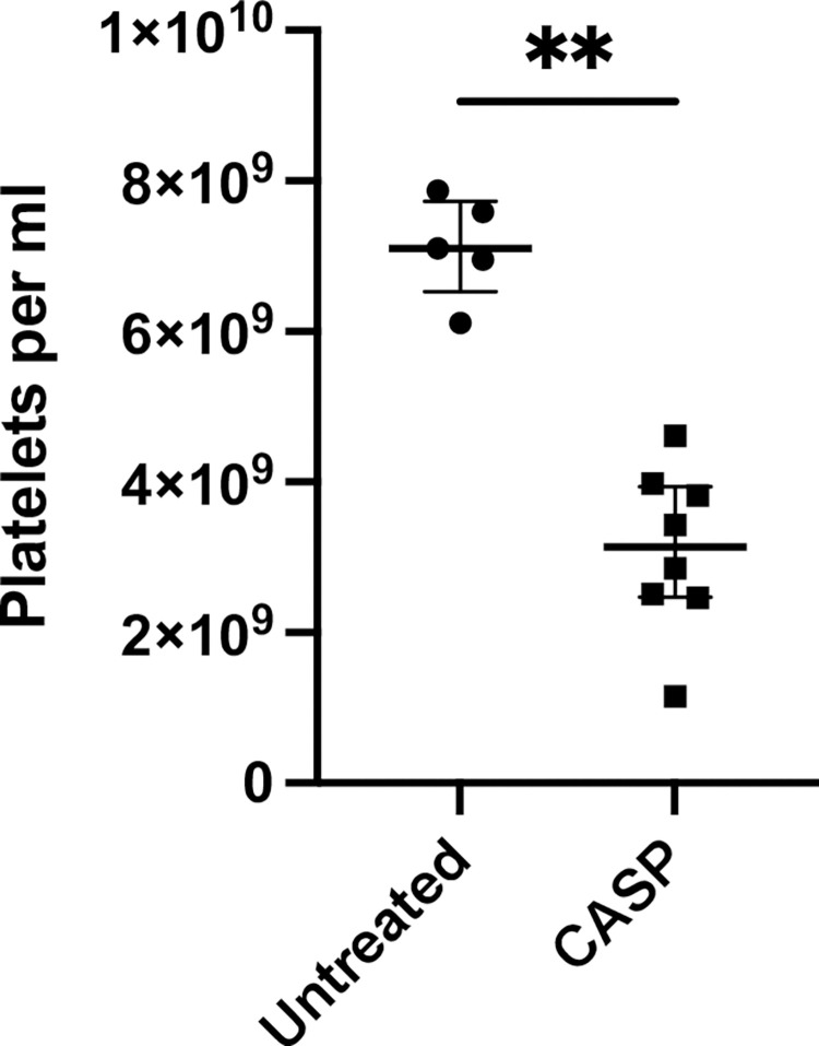
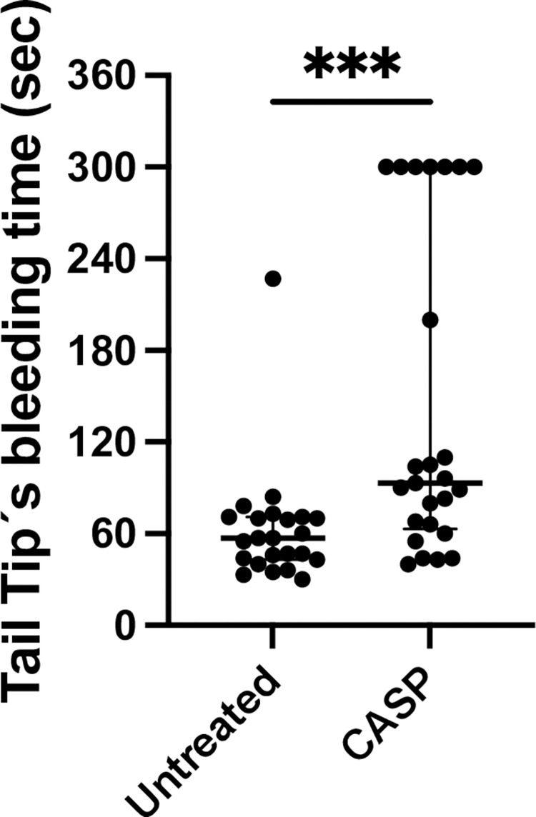
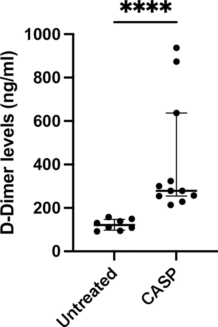
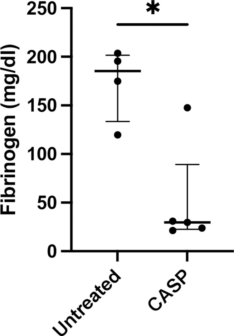
Background: Disseminated Intravascular Coagulation (DIC) is a life-threatening complication of sepsis. In surgical ICUs, DIC is frequently caused by abdominal sepsis, and the disarranged coagulation and complications often lead to death. The severity of sepsis is associated with a higher DIC score according to the parameters proposed by the International Society of Hemostasis and Thrombosis (ISTH) in 2001: platelet count, bleeding time (Quick), D-dimer, and fibrinogen. One problem in studying DIC is finding an adequate animal model that reflects the clinical situation of polymicrobial overwhelming infection.

Aims and methods: We investigated whether a well-established polymicrobial sepsis model of colon ascendens stent peritonitis (CASP) is suited to investigate the complexity of DIC. For this purpose, CASP-operated mice were examined 20 h after the operation with regard to coagulation parameters using cell counts, bleeding times, rotational thromboelastometry (ROTEM), ELISAs for D-dimer and fibrinogen, and platelet accumulation in affected organs via immunohistochemistry to see if the mice develop a coagulation disorder that meets the definition of DIC proposed by the ISTH 2001 consensus conference.

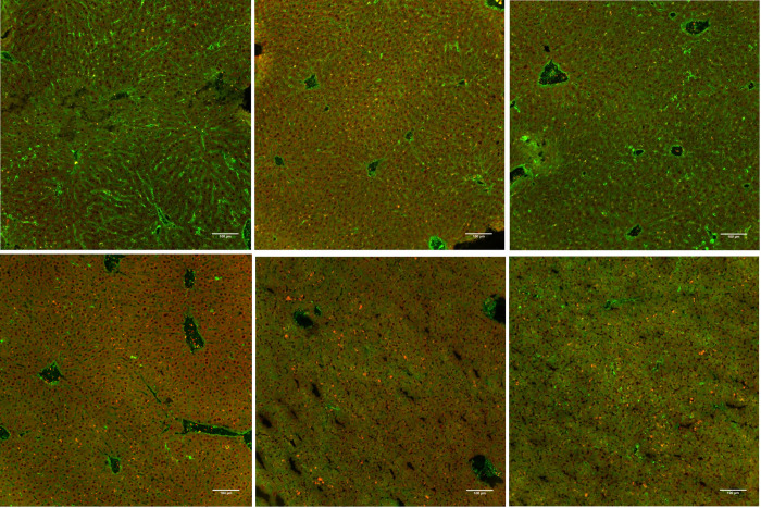
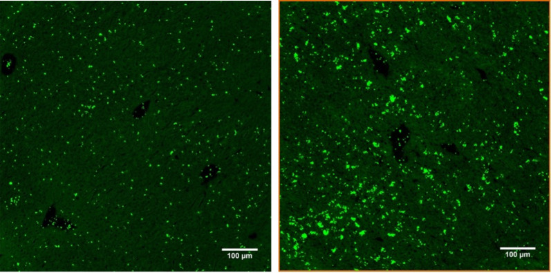
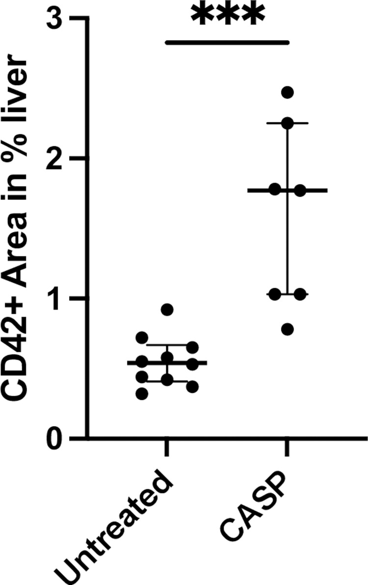
Results: Herein, we showed that the CASP model is an all-encompassing animal model to analyze the complexity of systemic DIC in murine abdominal sepsis. There is highly reproducible thrombocytopenia, a significant prolongation of the bleeding time, and a loss of fibrinogen in plasma. We also observed microvascular thrombosis due to platelet accumulation in the microcirculation of the liver.

Conclusion: The CASP model seems superior to other artificial models, e.g., injecting substances, for inducing DIC. CASP is one of the best true-to-life models for analyzing the complexity of disseminated intravascular coagulation in polymicrobial sepsis.


Conflict of interest statement

NO - The authors have declared that no competing interests exist.

Figures

Fig 1
Fig 1. Platelet count 20 hours after CASP surgery.
Untreated mice had a mean platelet count of 7.1 (1.76 IQR) × 109/ml, white column. Mice that underwent CASP surgery showed a reduction to 3.14 (3.46 IQR) × 109/ml, black column. Septic mice lost 60% of their platelets. The decrease in platelet count was highly significant (p = 0,0016**; n = 5 untreated group; n = 8 CASP group). The bold black line depicts the median.
Fig 2
Fig 2. Tail tip bleeding time in seconds.
Shown are bleeding times out of the tail tip of 8- to 10-week-old C57Bl/6 mice divided into CASP-operated, black column, and untreated animals, white column, 20 hours after CASP induction exhibiting significant differences in bleeding time (p < 0.001***; n = 23 untreated group; n = 25 CASP group). The tail tip bleeding time was 93 (260 IQR) seconds in septic mice and untreated mice showed a bleeding time of 57 (197 IQR) seconds. The bold black line depicts the median.
Fig 3
Fig 3. Clotting time (CT) extrinsic pathway rotational thromboelastometry ROTEM I.
Illustrated is the CT, measured via rotational thromboelastometry EXTEM in seconds of female 8- to 10-week-old C57Bl/6 mice untreated (white column) or 20 h after CASP surgery (CASP, black column); control mice (untreated) exhibited a stable clotting mechanism within 80 (21 IQR) seconds while 20 h after CASP surgery, the mice showed a prolongation of CT to 109 (409 IQR) seconds (p = 0.0043**; n = 6 untreated group; n = 5 CASP group). The bold black line depicts the median.
Fig 4
Fig 4. D-dimer levels in the blood of untreated or CASP-operated C57Bl/6 mice.
Statistical results of D-dimer levels 20 h after CASP surgery (CASP, black column) or untreated (white column) 8- to 10-week-old C57Bl/6. Basal blood levels of untreated animals 120.9 (66.2 IQR) ng/ml increased significantly to 278.6 (722.9 IQR) ng/ml in CASP-operated mice (p < 0.0001****; n = 8 untreated group; n = 11 CASP group); The bold black line depicts the median.
Fig 5
Fig 5. Fibrinogen blood levels 20 h after CASP surgery.
Statistical results of fibrinogen blood levels 20 h after CASP surgery in 8- to 10-week-old C57BL/6 mice depicting a significant decrease in fibrinogen plasma levels in CASP-operated mice (CASP, black column). Control animals (untreated, white column) showed 185.2 (83.91 IQR) mg/dl vs. 29.71 (126.3 IQR) mg/dl blood levels 20 h after CASP surgery (p = 0.03*; n = 4 untreated group; n = 5 CASP group). The bold black line depicts the median.
Fig 6
Fig 6. Frozen sections of the liver with immunohistochemistry staining with an anti-fibrinogen antibody Alexa 488 and platelet anti-CD 42c antibody TRITC.
Pictured: left row untreated control animals (a-c) showing liver sections of untreated 8- to 10-week-old C57Bl/6 (3 examples a-c untreated and d-f CASP), the green dye (Alexa 488) represents the fibrinogen amount in the liver sinusoids, the orange dye (TRITC) shows platelet aggregates, the right column is 20 h after CASP surgery (d-f, scale bar 100 μm, negative control not shown).
Fig 7
Fig 7. Immunohistochemical staining of frozen liver sections 20 h after CASP with anti-CD42c*FITC/Alexa 488.
Depicted are representative results of frozen sections from 8- to 10-week-old C57Bl/6 liver tissue with immunohistochemistry staining of platelets (green dye) with anti-CD 42c antibody*FITC/Alexa 488. The left row shows only a few platelets uniformly distributed in the liver tissue of untreated animals (a), while the right row shows platelet accumulation and aggregates in the liver tissue 20 h after CASP surgery (b); (scale bar 100 μm, negative control not shown, quantification of the r esults shown in Fig 8).
Fig 8
Fig 8. Accumulation of hepatic platelets 20 hours after CASP surgery.
Pictured is the percentage area of FITC/Alexa 488-positive areas on the fluorescent section in proportion to the entire section. There was a significant increase (p < 0.001***) in the percentage of the FITC/Alexa 488-positive area, i.e., an increased accumulation of hepatic platelets in CASP-operated animals of 1.77% (1.69% IQR) of the area (n = 7) compared to untreated animals with 0.54% (0.6% IQR) of the FITC-positive area (n = 10). The bold black line depicts the median.

References

    1. Iba T, Connors JM, Nagaoka I, Levy JH. Recent advances in the research and management of sepsis-associated DIC. Int J Hematol. 2021;113(1):24–33. doi: 10.1007/s12185-020-03053-y - DOI - PMC - PubMed
    1. Levi M. Another step in improving the diagnosis of disseminated intravascular coagulation in sepsis. Critical care (London, England). 2013;17(4):448. doi: 10.1186/cc12859 - DOI - PMC - PubMed
    1. Tang N, Li D, Wang X, Sun Z. Abnormal coagulation parameters are associated with poor prognosis in patients with novel coronavirus pneumonia. J Thromb Haemost. 2020;18(4):844–7. doi: 10.1111/jth.14768 - DOI - PMC - PubMed
    1. Akca S, Haji-Michael P, de Mendonça A, Suter P, Levi M, Vincent JL. Time course of platelet counts in critically ill patients. Crit Care Med. 2002;30(4):753–6. doi: 10.1097/00003246-200204000-00005 - DOI - PubMed
    1. Rice TW, Wheeler AP. Coagulopathy in critically ill patients: part 1: platelet disorders. Chest. 2009;136(6):1622–30. doi: 10.1378/chest.08-2534 - DOI - PubMed

Publication types