. 2022 Dec 9;378(6624):1111-1118.
doi: 10.1126/science.abq2787. Epub 2022 Dec 8.

Tuberculosis treatment failure associated with evolution of antibiotic resilience

Affiliations

Tuberculosis treatment failure associated with evolution of antibiotic resilience

Qingyun Liu et al. Science. .

Abstract

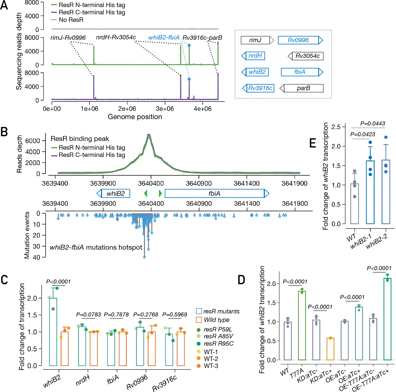
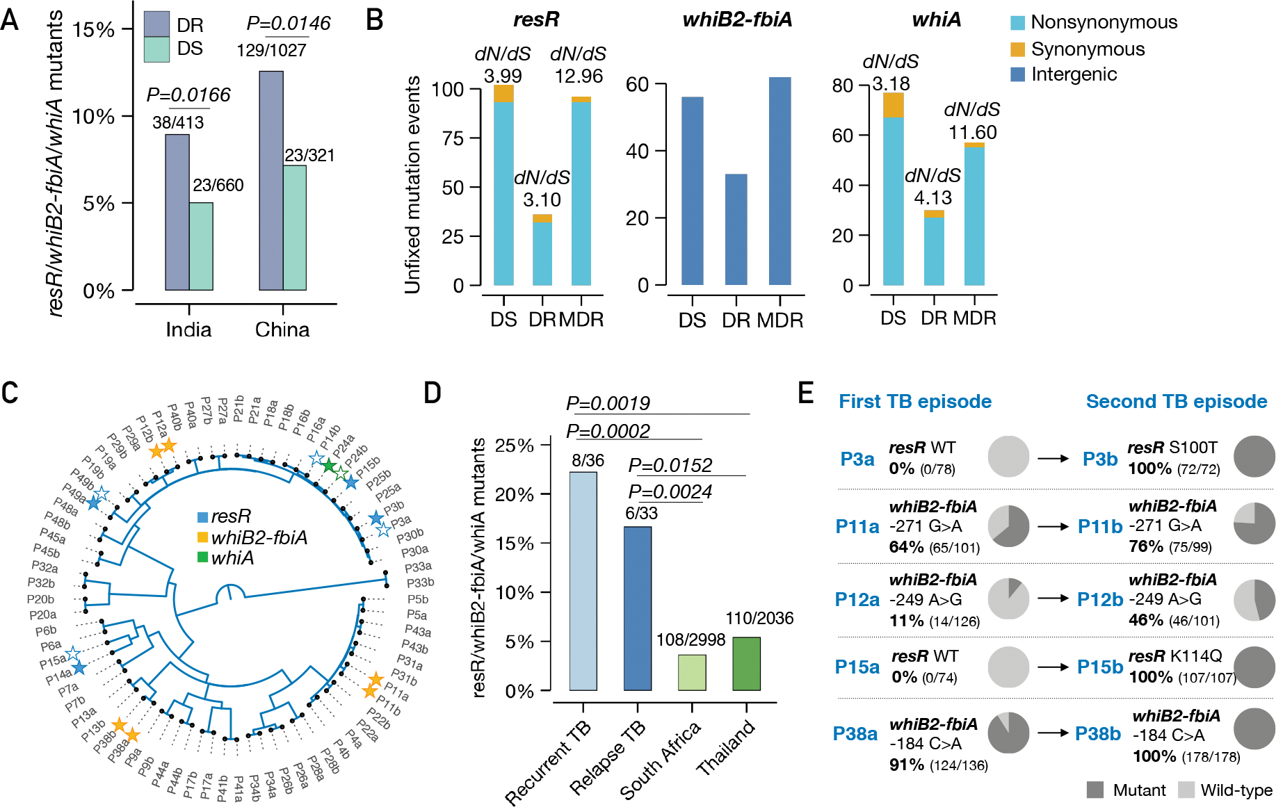
The widespread use of antibiotics has placed bacterial pathogens under intense pressure to evolve new survival mechanisms. Genomic analysis of 51,229 Mycobacterium tuberculosis (Mtb)clinical isolates has identified an essential transcriptional regulator, Rv1830, herein called resR for resilience regulator, as a frequent target of positive (adaptive) selection. resR mutants do not show canonical drug resistance or drug tolerance but instead shorten the post-antibiotic effect, meaning that they enable Mtb to resume growth after drug exposure substantially faster than wild-type strains. We refer to this phenotype as antibiotic resilience. ResR acts in a regulatory cascade with other transcription factors controlling cell growth and division, which are also under positive selection in clinical isolates of Mtb. Mutations of these genes are associated with treatment failure and the acquisition of canonical drug resistance.


Conflict of interest statement

Competing interests: The authors declare no competing interests.

Figures

Fig. 1.
Fig. 1.. Ongoing positive selection in the global Mtb population.
(A) Diagram of fixed and unfixed variants. (B) Genes and IGRs with signal of ongoing positive selection. Known drug-resistance (DR) or drug-tolerance (DT) genes are marked with capsule symbols. Red stars indicate transcriptional regulators. The map shows the geographic origin of the clinical Mtb isolates and their DR status; DT (drug-tolerance), MDR (multi-drug resistance). (C) Categories of genes under positive selection, with adjusted P values for enrichment. (D) dN/dS ratio of resR in DR and DS strains, P value by unpaired t test. (E) Structural model of the ResR dimer (cyan, predicted by Alphafold) aligned to MerR-family homolog CueR (orange) (49) in complex with duplex DNA with common ResR clinical mutations indicated (yellow). (F) Schematic of construction of mutations in the chromosomal copy of resR in Mtb.
Fig. 2.
Fig. 2.. resR mutants showed faster recovery after drug exposure.
(A) MICs of resR mutants and wild type for 8 anti-TB drugs. INH: Isoniazid; RIF: Rifampicin; OFX: Ofloxacin; ETH: Ethionamide; EMB: Ethambutol; DCS: D-cycloserine; BDQ: Bedaquiline; LZD: Linezolid. Drug-resistance breakpoint is the drug concentration that defines clinical drug resistance. (B) Time-kill kinetics for 8 different drugs for resR mutant and wild-type Mtb strains. The concentration of each drug was 100-fold MIC. * symbol indicates statistical significance (P < 0.05) by unparied t test. (C) Representative images illustrating the post-antibiotic recovery dynamics of resR mutant (R95C) and wild-type (WT) Mtb strains.
Fig. 3.
Fig. 3.. Quantitative colony-size tracking indicated shortened PAE in resR mutants.
(A) Quantitative colony-size tracking in pixel unit (px.) for resR mutant and wild-type Mtb strains in the presence or absence of antibiotic exposure (24h). (B) Duration from plating to the appearance of visible colonies (Time to colony formation) for resR mutant and wild-type strains after exposure to indicated antibiotics (24h) or no drug exposure (None). P=0.7291 for no drug exposure group and P<0.0001 for all drug groups, by Mann-Whitney U test. (C) A bar plot depicting the median values of post-antibiotic delay of wild type (light green) and resR mutants (dark green).
Fig. 4.
Fig. 4.. Antibiotic resilience characterized by more rapid resumption of cell wall synthesis.
(A) A schematic diagram of pulse-chase experiment for tracking the regrowth of Mtb cells post antibiotic exposure using fluorescent d-amino acid incorporation (NADA and HADA). (B) Representative microscopy snapshots of the resR mutants and wild type during regrowth after 24 hours of INH (100-fold MIC) exposure (the upper panel) and no antibiotic exposure (the lower panel). Scale bar: 10 m. (C) Quantitative comparison of outgrowth between resR mutant and wild type in post antibiotic exposure group and no antibiotic exposure group, P<0.0001 for 24h and 36h of post antibiotic exposure group by double-sided Kolmogorov–Smirnov test. (D) Flow cytometry for NADA incorporation into Mtb cells after 24 hours recovery post antibiotic exposure or without drug exposure.
Fig. 5.
Fig. 5.. ResR activates whiB2 and clinical mutations lead to upregulation of whiB2.
(A) IDAP-Seq identified binding sites of ResR. (B) ResR binding peaks overlaid with the mutations identified in clinical strains between whiB2-fbiA. The transcriptional start sites of whiB2 and fbiA are annotated (green arrows). (C) Ford change of transcription of putative ResR targets in resR mutants and wild-type strains, P values by the Wald test implemented in DEseq2. (D) Transcriptional changes of whiB2 in M. smegmatis strains: wild-type (WT), resR point mutant (T77A), CRISPR-i knock-down of resR (KD), merodiploid overexpression of wild-type resR (OE) or T77A mutant form (OE-T77A). The absence or presence of aTc was specified by (−) or (+), P values given by unpaired t test. (E) whiB2-fbiA mutant (3640375 C>T) exhibited upregulation of whiB2. whiB2–1 and whiB2–2 refer to the two parallel mutants, and P values given by unpaired t test.
Fig. 6.
Fig. 6.. resR, whiB2-fbiA and whiA mutants were associated with canonical drug resistance and relapse of drug-susceptible tuberculosis.
(A) The proportion of resR, whiB2-fbiA and whiA mutants in DR and DS strains sequenced from India and China, P values by Fisher’s exact test. (B) Unfixed mutations in DS, DR and MDR (resistant to RIF and INH) strains. (C) A phylogenetic tree of paired Mtb isolates from 36 recurrent TB patients. Solid stars indicate isolates in which mutations were detected while empty stars indicate absence of mutations in one of the paired isolates. (D) Percentage of isolates with mutations in resR/whiB2-fbiA/whiA. “Recurrent TB” includes 3 patients with Mtb isolates suggestive of re-infection. (E) Mutational trajectory in Mtb isolates from the first TB to second TB episodes in 5 pairs of isolates. The mutations in other three pairs: P14 (resR, D144N; 100% in P14a, 0% in P14b); P49 (resR, R95H; 8.7% in P49a, 0% in P49b); P24 (whiA, A131T; 11.6% in P24a, 0% in P24b).

References

    1. Fisher RA, Gollan B, Helaine S, Persistent bacterial infections and persister cells. Nat Rev Microbiol 15, 453–464 (2017) (10.1038/nrmicro.2017.42). - DOI - PubMed
    1. Liu J, Gefen O, Ronin I, Bar-Meir M, Balaban NQ, Effect of tolerance on the evolution of antibiotic resistance under drug combinations. Science 367, 200–204 (2020) (10.1126/science.aay3041). - DOI - PubMed
    1. Harms A, Maisonneuve E, Gerdes K, Mechanisms of bacterial persistence during stress and antibiotic exposure. Science 354, aaf4268 (2016) (10.1126/science.aaf4268). - DOI - PubMed
    1. Safi H et al., Phase variation in Mycobacterium tuberculosis glpK produces transiently heritable drug tolerance. Proceedings of the National Academy of Sciences of the United States of America 116, 19665–19674 (2019) (10.1073/pnas.1907631116). - DOI - PMC - PubMed
    1. Murray JF, Schraufnagel DE, Hopewell PC, Treatment of Tuberculosis. A Historical Perspective. Ann Am Thorac Soc 12, 1749–1759 (2015) (10.1513/AnnalsATS.201509-632PS). - DOI - PubMed

MeSH terms