Review
. 2023 Jan:176:105948.
doi: 10.1016/j.nbd.2022.105948. Epub 2022 Dec 5.

Brain edema formation and therapy after intracerebral hemorrhage

Affiliations
Review

Brain edema formation and therapy after intracerebral hemorrhage

Yingfeng Wan et al. Neurobiol Dis. 2023 Jan.

Abstract

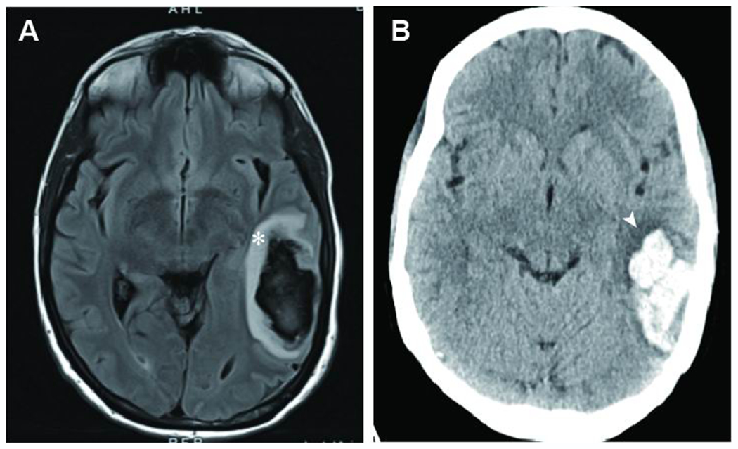
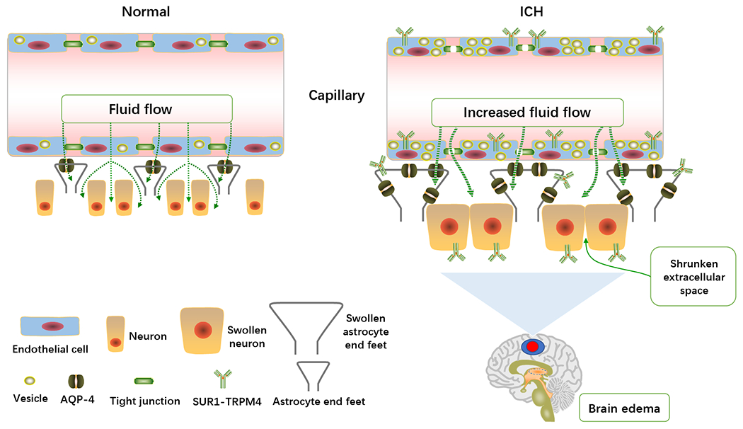
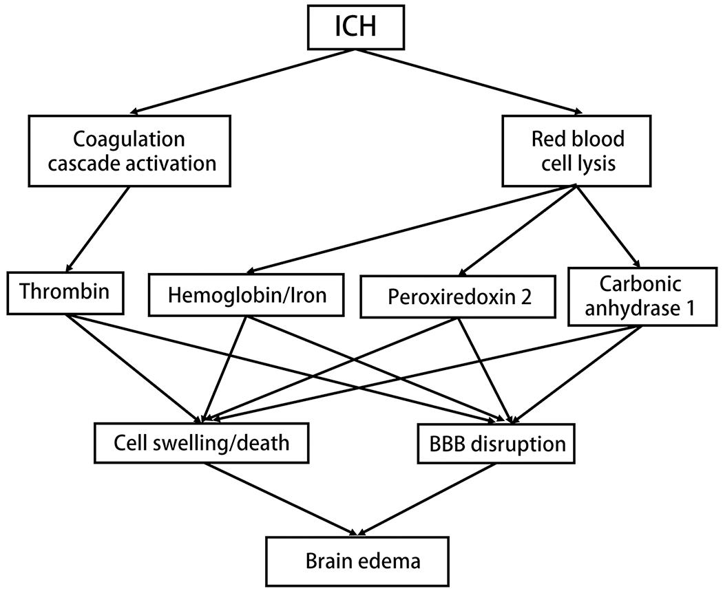
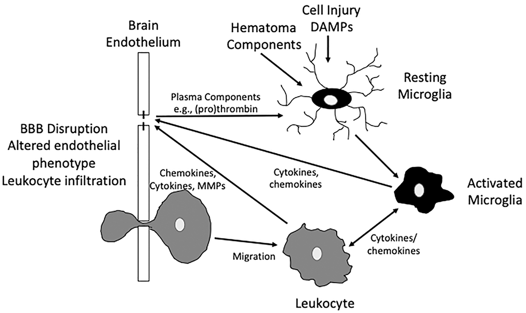
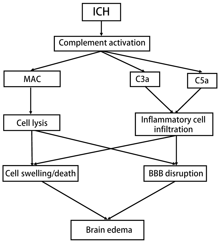
Intracerebral hemorrhage (ICH) accounts for about 10% of all strokes in the United States of America causing a high degree of disability and mortality. There is initial (primary) brain injury due to the mechanical disruption caused by the hematoma. There is then secondary injury, triggered by the initial injury but also the release of various clot-derived factors (e.g., thrombin and hemoglobin). ICH alters brain fluid homeostasis. Apart from the initial hematoma mass, ICH causes blood-brain barrier disruption and parenchymal cell swelling, which result in brain edema and intracranial hypertension affecting patient prognosis. Reducing brain edema is a critical part of post-ICH care. However, there are limited effective treatment methods for reducing perihematomal cerebral edema and intracranial pressure in ICH. This review discusses the mechanisms underlying perihematomal brain edema formation, the effects of sex and age, as well as how edema is resolved. It examines progress in pharmacotherapy, particularly focusing on drugs which have been or are currently being investigated in clinical trials.

Keywords: Brain edema; Intracerebral hemorrhage; Mechanisms; Therapy.


Conflict of interest statement

Declaration of Competing Interest Yingfeng Wan, Katherine G Holste, Ya Hua, Richard F. Keep, and Guohua Xi declare no conflict of interests.

Figures

Figure 1:
Figure 1:
(A) One ICH patient’s 2 days MRI FLAIR image after hemorrhage showing significant hyperintense perihematomal edema area (indicated by white asterisk) and compression of the lateral ventricle. (B) The same ICH patient’s 1 hour CT image after hemorrhage showing low-density brain edema region (indicated by white arrow) around the hematoma.
Figure 2:
Figure 2:. Schematic diagram of fluid movement and relevant mechanisms in normal and ICH brain.
ICH induced greater AQP4 channel opening in the end feet of astrocyte and SUR1-TRPM4 expression in the BBB endothelial cells, astrocytes and neurons. Swollen neuron and astrocyte end feet were observed after ICH. It can be noticed that tight junction disruption and shrunken extracellular space after ICH, increased fluid flow into brain parenchyma and more vesicles were found in endothelial cells.
Figure 3:
Figure 3:. Hematoma components and brain edema.
Erythrolysis leads to release of hemoglobin, PRX-2 and CA-1, which contribute to perihematomal edema after ICH. Thrombin formation after coagulation cascade activation also contributes to brain edema.
Figure 4:
Figure 4:. Inflammation and brain edema.
Inflammation after ICH is triggered by cell injury with the release of DAMPs and the release of different molecules from the hematoma (e.g., thrombin, Hb and PRX2). There is then an inflammatory cascade involving microglia activation, BBB disruption and leukocyte (neutrophils, macrophages, lymphocytes and NK cells) infiltration into brain with a complex interplay between these cells. That interplay involves a range of signaling molecules (cytokines, chemokines, MMPs – see text) as well as other cell types including perivascular macrophages and astrocytes (omitted for simplicity).
Figure 5:
Figure 5:. Complement activation and brain edema following ICH.
MAC forms pores on cell membrane contributing to cerebral edema through red blood cell lysis, brain cell swelling/death and BBB disruption. C3a and C5a lead to chemotaxis of circulating inflammatory cells after ICH and cause perihematomal brain edema.

Similar articles

Cited by

References

    1. Abbasi V, et al., 2017. The effect of acetazolamide on intracerebral hemorrhage in stoke patients. Int. J. Adv. Med 4, 148–151.
    1. Akk A, et al., 2019. Complement activation on neutrophils initiates endothelial adhesion and extravasation. Mol Immunol. 114, 629–642. - PMC - PubMed
    1. Al-Shahi Salman R, et al., 2018. Absolute risk and predictors of the growth of acute spontaneous intracerebral haemorrhage: a systematic review and meta-analysis of individual patient data. Lancet Neurol. 17, 885–894. - PMC - PubMed
    1. Amit Y, Brenner T, 1993. Age-dependent sensitivity of cultured rat glial cells to bilirubin toxicity. Exp Neurol. 121, 248–55. - PubMed
    1. Anderson CS, et al., 2010. Effects of early intensive blood pressure-lowering treatment on the growth of hematoma and perihematomal edema in acute intracerebral hemorrhage: the Intensive Blood Pressure Reduction in Acute Cerebral Haemorrhage Trial (INTERACT). Stroke. 41, 307–12. - PubMed

Publication types