

Micro-Computed Tomography (µCT) as a Tool for High-Resolution 3D Imaging and Analysis of Intraocular Lenses: Feasibility and Proof of the Methodology to Evaluate YAG Pits
- PMID: 36481844
- PMCID: PMC9834457
- DOI: 10.1007/s40123-022-00622-8
Micro-Computed Tomography (µCT) as a Tool for High-Resolution 3D Imaging and Analysis of Intraocular Lenses: Feasibility and Proof of the Methodology to Evaluate YAG Pits
Abstract
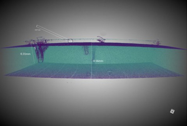
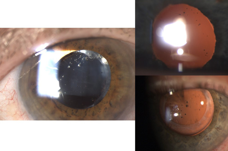
Introduction: Posterior capsule opacification (PCO) is the most frequent late sequelae after successful cataract surgery. Neodymium:yttrium aluminum garnet (Nd:YAG) laser capsulotomy is considered the gold standard and a well-accepted, safe, and effective measure in treating PCO. However, iatrogenic damage of the intraocular lens (IOL) due to inappropriate focusing is a quite common side effect. These permanent defects (YAG pits) can critically affect overall optical quality.
Methods: In this laboratory study, we used the micro-computed tomography (µCT) technique to obtain high-resolution 3D images of the lens and the YAG pits.
Results: To the best of our knowledge, this is the first description of a detailed analysis of IOLs with µCT technology. This non-destructive technique seems to be ideal for comparative studies, measuring dimensions of the damage, and visualizing shooting channels within the material.
Conclusion: µCT is excellently suited to examine an IOL in detail, analyze optics and haptics in three dimensions, and to describe all kinds of changes within the IOL without damaging it.
Keywords: Damage in intraocular lenses; Laboratory study to evaluate IOL quality; Micro-computed tomography (µCT); New technique; YAG pits.
© 2022. The Author(s).
Figures
Similar articles
-
Incorrectly Focused Neodymium:Yttrium-Aluminum-Garnet (Nd:YAG) Laser Beam Leads to Massive Destructive Effects in Small-Aperture (Pinhole) Intraocular Lenses.Ophthalmol Ther. 2024 Oct;13(10):2745-2758. doi: 10.1007/s40123-024-01007-9. Epub 2024 Aug 17. Ophthalmol Ther. 2024. PMID: 39153117 Free PMC article.
-
Evaluating impact of Nd: YAG laser associated defects on optical quality of hydrophilic and hydrophobic intraocular lenses using visualization of light propagation and USAF test targets.BMC Ophthalmol. 2022 Dec 16;22(1):494. doi: 10.1186/s12886-022-02738-8. BMC Ophthalmol. 2022. PMID: 36527032 Free PMC article.
-
A Novel 3D High Resolution Imaging Method Using Correlative X-Ray and Electron Microscopy to Study Neodymium-Doped Yttrium Aluminum Garnet Laser-Induced Defects in Intraocular Lenses.Ophthalmic Res. 2024;67(1):292-300. doi: 10.1159/000539243. Epub 2024 May 8. Ophthalmic Res. 2024. PMID: 38718759
-
Image Contrast and Spectral Transmission in Intraocular Lenses with Nd:YAG Pits.Curr Eye Res. 2023 Oct;48(10):911-918. doi: 10.1080/02713683.2023.2229540. Epub 2023 Jun 29. Curr Eye Res. 2023. PMID: 37382106
-
Posterior Capsule Opacification after Cataract Surgery via Implantation with Hydrophobic Acrylic Lens Compared with Silicone Intraocular Lens: A Systematic Review and Meta-Analysis.J Ophthalmol. 2022 Feb 25;2022:3570399. doi: 10.1155/2022/3570399. eCollection 2022. J Ophthalmol. 2022. PMID: 35251708 Free PMC article. Review.
Cited by
-
Nano-Indentation to Determine Mechanical Properties of Intraocular Lenses: Evaluating Penetration Depth, Material Stiffness, and Elastic Moduli.Ophthalmol Ther. 2023 Aug;12(4):2087-2101. doi: 10.1007/s40123-023-00728-7. Epub 2023 May 21. Ophthalmol Ther. 2023. PMID: 37211587 Free PMC article.
-
Precise Posterior Nd:YAG Capsulotomy without Creating Defects is Key for Patients' Quality of Vision.Clin Ophthalmol. 2024 Feb 5;18:361-364. doi: 10.2147/OPTH.S451238. eCollection 2024. Clin Ophthalmol. 2024. PMID: 38343902 Free PMC article. No abstract available.
-
Incorrectly Focused Neodymium:Yttrium-Aluminum-Garnet (Nd:YAG) Laser Beam Leads to Massive Destructive Effects in Small-Aperture (Pinhole) Intraocular Lenses.Ophthalmol Ther. 2024 Oct;13(10):2745-2758. doi: 10.1007/s40123-024-01007-9. Epub 2024 Aug 17. Ophthalmol Ther. 2024. PMID: 39153117 Free PMC article.
-
Evaluating impact of Nd: YAG laser associated defects on optical quality of hydrophilic and hydrophobic intraocular lenses using visualization of light propagation and USAF test targets.BMC Ophthalmol. 2022 Dec 16;22(1):494. doi: 10.1186/s12886-022-02738-8. BMC Ophthalmol. 2022. PMID: 36527032 Free PMC article.
References
-
- Market Scope. 2020 IOL market report: mid-year update. Market Scope, St. Louis, MO. https://www.market-scope.com. Accessed 28 Aug 2020.
-
- Rönbeck M, Lundström M, Kugelberg M. Study of possible predictors associated with self-assessed visual function after cataract surgery. Ophthalmology. 2011;118(9):1732–8. 10.1016/j.ophtha.2011.04.013. (Erratum in: Ophthalmology. 2011118(10):1965). - PubMed
LinkOut - more resources
Full Text Sources
