. 2023 Apr;46(4):959-971.
doi: 10.1038/s41440-022-01094-w. Epub 2022 Dec 9.

Mapping the protein binding site of the (pro)renin receptor using in silico 3D structural analysis

Affiliations

Mapping the protein binding site of the (pro)renin receptor using in silico 3D structural analysis

Akio Ebihara et al. Hypertens Res. 2023 Apr.

Erratum in

Abstract

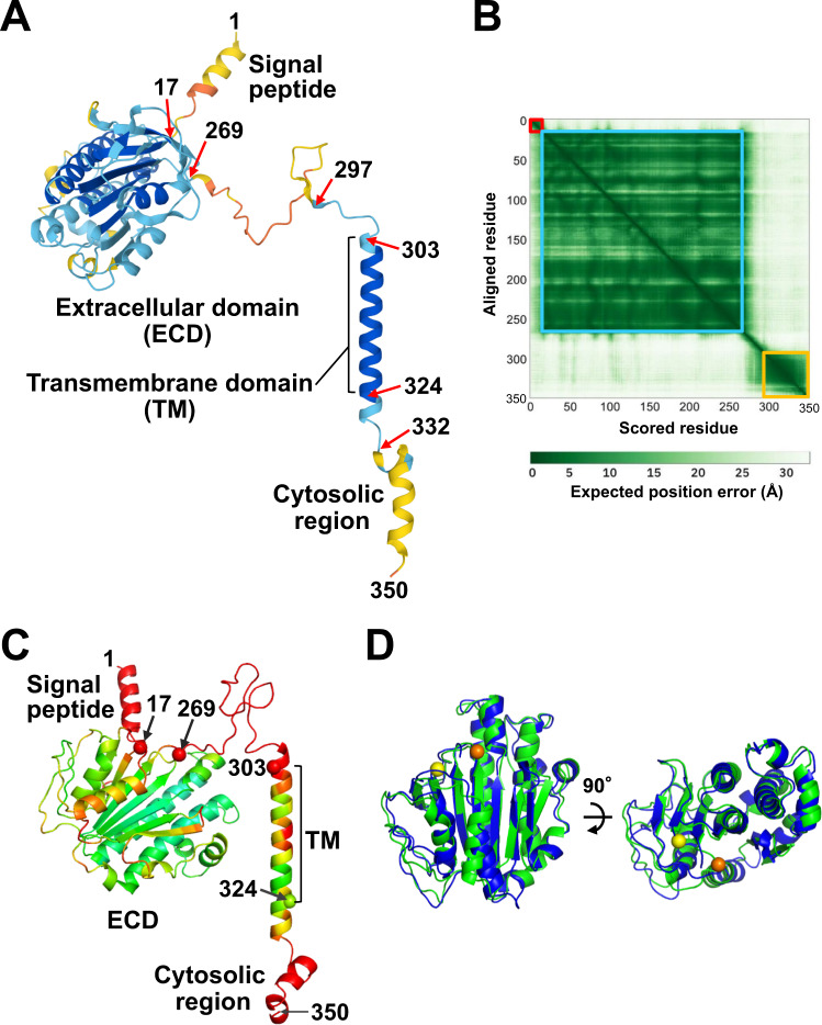
We have previously reported that monoclonal antibodies against the (pro)renin receptor [(P)RR] can reduce the Wnt/β-catenin-dependent development of pancreatic ductal adenocarcinoma (PDAC), the most common pancreatic cancer. Antibodies against two (P)RR regions (residues 47-60 and 200-213) located in the extracellular domain (ECD) reduced the proliferation of human PDAC cells in vitro. Although these regions probably participate in the activation of Wnt/β-catenin signaling, their functional significance remains unclear. Moreover, the (P)RR ECD is predicted to possess an intrinsically disordered region (IDR), which allows multiple protein interactions because of its conformational flexibility. In this study, we investigated the significance of the two regions and the IDR by in silico 3D structural analysis using the AlphaFold2 program and evolutionary sequence conservation profile. The model showed that ECD adopted a folded domain (residues 17-269) and had an IDR (residues 270-296). The two regions mapped onto the structural model formed a continuous surface patch comprising evolutionarily conserved hydrophobic residues. The homodimeric structure predicted by AlphaFold2 showed that full-length (P)RR comprising the ECD, single-span transmembrane, and cytoplasmic domains formed a twofold symmetric dimer via the ECD, which explains the experimentally proven homodimerization. The dimer model possessed two hand-shaped grooves with residues 47-60 and 200-213 in their palms and the IDR as their fingers. Based on these findings, we propose that the IDR-containing hydrophobic grooves act as a binding site for (P)RR and perform multiple functions, including Wnt signaling activation. Antibodies against the (pro)renin receptor residues 47-60 and 200-213 can inhibit pancreatic ductal adenocarcinoma (PDAC) cell proliferation by suppressing Wnt signaling. This study provides 3D structural insights into receptor binding and one-to-many interactions, which underpin the functional versatility of this receptor.

Keywords: (Pro)renin receptor; AlphaFold2; Intrinsically disordered region; Pancreatic ductal adenocarcinoma; Wnt/β-catenin signaling.


Conflict of interest statement

The authors declare no competing interests.

Figures

None
Antibodies against the (pro)renin receptor residues 47–60 and 200–213 can inhibit pancreatic ductal adenocarcinoma (PDAC) cell proliferation by suppressing Wnt signaling. This study provides 3D structural insights into receptor binding and one-to-many interactions, which underpin the functional versatility of this receptor.
Fig. 1
Fig. 1
Predicted structure of human (P)RR. A Cartoon representation of the AF2 structural model. The model includes the signal peptide (residues 1–16), extracellular domain (ECD; residues 17–302), transmembrane domain (TM; residues 303–323), and cytosolic region (residues 324–350). The structure is colored according to the predicted local-distance difference test (pLDDT) score in blue, cyan, yellow, and orange for the regions that were most confidently predicted (pLDDT > 90), confidently predicted (90 > pLDDT >70), predicted with low confidence (70 > pLDDT >50), and predicted with very low confidence (pLDDT < 50), respectively. B Predicted aligned error (PAE) plot. The PAE values of residues 1–16, 17–269, and 290–350 are illustrated with overlaid squares in red, cyan, and yellow, respectively. C Cartoon representation of the RoseTTAFold structural model. The structure is colored according to the pLDDT: blue for the most confidently predicted regions (pLDDT ≥ 0.9) and red for those predicted with very low confidence (pLDDT ≤ 0.5) based on a color spectrum of red, yellow, green, cyan, and blue. D Secondary structure-based superimposition of RoseTTAFold ECD (green) on AF2 ECD (blue). Cα atoms of residues 17 and 269 of the RoseTTAFold model are shown as yellow and orange spheres, respectively. The right view is the same representation rotated by 90°
Fig. 2
Fig. 2
Structural similarity of human (P)RR to alkaline phosphatase (ALP) family proteins. A The overall structure of the ECD with a cartoon representation (α-helices shown in red and β-strands in yellow). The Cα atom of residue 17 is shown as a blue sphere, and that of residue 269 is shown as an orange sphere. B Structure-based superimposition of human (P)RR ECD (green) on PhoK (blue; PDB ID: 5XWK, residues 31–560). Cα traces of both proteins are shown as ribbons. C PhoK active site. Active site residues are shown as sticks. Two bound zinc centers (purple) and a phosphate ion (oxygen atom, red; phosphorus atom, orange) are shown as spheres. D The human (P)RR residues that occupy spatially equivalent positions in the ALP active site. Amino acid residues that may bind to a metal and a phosphate ion are shown in stick representation
Fig. 3
Fig. 3
Multiple sequence alignment of human (P)RR and its sequence homologs. The sequence homologs are specified with clade/species names (common name and UniRef cluster ID): Boreoeutheria (mammal; UniRef90_P81134), Crocodylia (alligator; UniRef90_A0A151NQQ9), Neognathae (bird; UniRef90_H0Z8C1), Latimeria chalumnae (coelacanth; UniRef90_H3AI83), Xenopus (clawed frog, UniRef90_Q5M8F1), Percomorphaceae (ray-finned fish; UniRef90_A0A4W6D9M0), Branchiostoma (Amphioxus; UniRef90_C3YJH1), and Stichopus japonicus (sea cucumber; UniRef90_A0A2G8L3G6). Identity (%) represents the percentage sequence identity to human (P)RR. Identical residues are shown as white characters with a red background and similar residues with red characters with a white background. The secondary structures of the human (P)RR AF2 model are shown at the top: α, α-helix; β, β-strand; η, coil. The processing site of site-1 protease (S1P) is depicted as a black triangle
Fig. 4
Fig. 4
Molecular surface properties of human (P)RR. A Surface representation of two regions involved in the antiproliferative effect against PDAC: residues 47–60 (red) and 200–213 (blue). The TM is colored yellow. B Evolutionary conservation profile shown in surface representation. Each amino acid residue is colored according to its ConSurf conservation score. The color-coding bar shows the ConSurf coloring scheme, which varies from green (highly variable; score 1) to purple (highly conserved; score 9). C Electrostatic surface potential colored from negative (−5 kT/e, red) to positive (+5 kT/e, blue). D Surface representation colored by hydrophobicity with a color gradient of red (most hydrophobic) to white (least hydrophobic). E Human (P)RR monomer depicted as a gray transparent surface with a cartoon representation (α-helices shown in red and β-strands in yellow). F (Middle) Another conservation profile shown in surface representation. Residues 47–60 and 200–213 are shown in red and blue, respectively. (Left and right) Two views are shown of the same representation related by a 90° rotation
Fig. 5
Fig. 5
Analysis of predicted homodimeric structures of human (P)RR. A Two chains of the ECD (residues 17–270) are depicted as a gray transparent surface with a cartoon representation. Each chain is colored on the rainbow scale from the N-terminus (blue) to the C-terminus (red). B Two chains of the full-length human (P)RR (residues 17–350) in a cartoon representation. Each chain is colored in rainbow format, except yellow, which indicates TM regions. C Surface representation of the full-length human (P)RR homodimer with the two chains colored gray and pale cyan. Residues 47–60, 200–213, and 281 are shown in red, blue, and green, respectively. D Evolutionary conservation profile of human (P)RR evaluated in terms of local structural environment. The Consurf scores are shown as dots, which are color-coded according to the local structural environment: solvent accessible, blue; solvent inaccessible, red; dimer interface, green. E Regionwise averaged Consurf scores of human (P)RR. The region “others” contains all ECD residues except 47–60, 105–153, 200–213, and 270–296. F (P)RR homodimer viewed perpendicular to the twofold axis. One chain is drawn in blue and the other in cyan. Residues 105–153 are colored red. The dimer interface residues are represented by stick models (green). G A close-up of solvent-accessible highly conserved residues (green) in two regions: residues 47–60 (red) and 200–213 (blue)
Fig. 6
Fig. 6
Mapping the protein binding sites of (P)RR. A Surface representation of the ECD of the full-length human (P)RR homodimer. Palm, residues 47–60 (blue) and 200–213 (red); thumb, residues 65–70 (red dotted); and fingers, residues 270–296 (blue botted). The model is shown with the same color coding as in Fig. 5C, except the IDR is shown in pink (residues 270–296). Surface representations with hydrophobicity score (B) and conservation profile (C) are shown in the same orientation as in (A). D Proposed “catch and tether” mechanism of (P)RR. (P)RR catches a loop with its “hand” and tethers two proteins. E AF2 structural models of FZD8, LRP6, and (P)RR depicted on the same scale for comparison. CRD cysteine-rich domain, E1–E4 four β-propeller/epidermal growth factor repeats, L1–L3 three low-density lipoprotein receptor type A repeats. The LRP6 structure downstream from L3 is shown in cartoon representation

Similar articles

Cited by

References

    1. Nguyen G, Delarue F, Burcklé C, Bouzhir L, Giller T, Sraer JD. Pivotal role of the renin/prorenin receptor in angiotensin II production and cellular responses to renin. J Clin Invest. 2002;109:1417–27. doi: 10.1172/JCI0214276. - DOI - PMC - PubMed
    1. Ichihara A, Yatabe MS. The (pro)renin receptor in health and disease. Nat Rev Nephrol. 2019;15:693–712. doi: 10.1038/s41581-019-0160-5. - DOI - PubMed
    1. Shibayama Y, Takahashi K, Yamaguchi H, Yasuda J, Yamazaki D, Rahman A, et al. Aberrant (pro)renin receptor expression induces genomic instability in pancreatic ductal adenocarcinoma through upregulation of SMARCA5/SNF2H. Commun Biol. 2020;3:724. doi: 10.1038/s42003-020-01434-x. - DOI - PMC - PubMed
    1. Peters J. The (pro)renin receptor and its interaction partners. Pflug Arch. 2017;469:1245–56. doi: 10.1007/s00424-017-2005-z. - DOI - PubMed
    1. Kanda A, Ishida S. (Pro)renin receptor: involvement in diabetic retinopathy and development of molecular targeted therapy. J Diabetes Investig. 2019;10:6–17. doi: 10.1111/jdi.12842. - DOI - PMC - PubMed