. 2022 Dec 8;5(1):1349.
doi: 10.1038/s42003-022-04305-9.

Controlled release of growth factors using synthetic glycosaminoglycans in a modular macroporous scaffold for tissue regeneration

Affiliations

Controlled release of growth factors using synthetic glycosaminoglycans in a modular macroporous scaffold for tissue regeneration

Z Söderlund et al. Commun Biol. .

Abstract

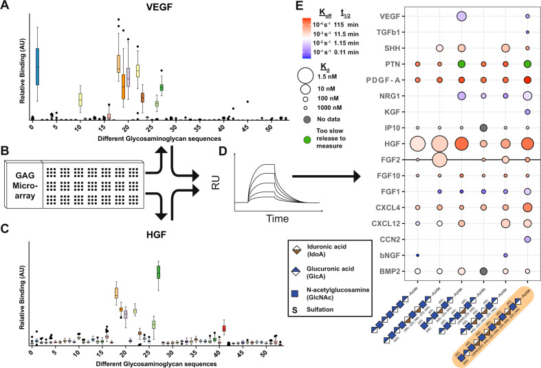
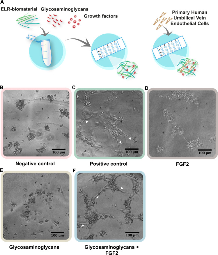
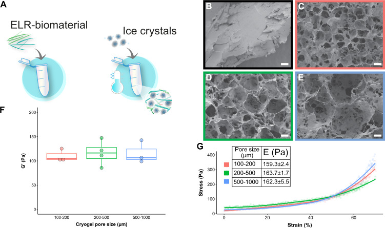
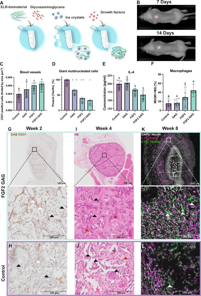
Healthy regeneration of tissue relies on a well-orchestrated release of growth factors. Herein, we show the use of synthetic glycosaminoglycans for controlled binding and release of growth factors to induce a desired cellular response. First, we screened glycosaminoglycans with growth factors of interest to determine kon (association rate constant), koff (dissociation rate constant), and Kd (equilibrium rate constant). As proof-of-concept, we functionalized an elastin-like recombinamer (ELR) hydrogel with a synthetic glycosaminoglycan and immobilized fibroblast growth factor 2 (FGF2), demonstrating that human umbilical vein endothelial cells cultured on top of ELR hydrogel differentiated into tube-like structures. Taking this concept further, we developed a tunable macroporous ELR cryogel material, containing a synthetic glycosaminoglycan and FGF2 that showed increased blood vessel formation and reduced immune response compared to control when implanted in a subcutaneous mouse model. These results demonstrated the possibility for specific release of desired growth factors in/from a modular 3D scaffold in vitro and in vivo.


Conflict of interest statement

The authors declare the following competing interests: J.L. is founder of Glycan Therapeutics LLC. The remaining authors declare no competing interests.

Figures

Fig. 1
Fig. 1. Screening of growth factor interactions with synthetic glycosaminoglycans.
A Relative binding of VEGF to 52 different synthetic glycosaminoglycans measured on a microarray. B An illustration of the microarray. C Relative binding of HGF in the microarray to show the difference in binding pattern depending on the glycosaminoglycan. For A and C data is shown in a box and whisker (as Tukey) of 36 binding spots for each synthetic glycosaminoglycan. D Six synthetic glycosaminoglycans with different binding patterns were chosen for further analyses using surface plasmon resonance (SPR) to determine their binding in a quantitative way compared to the semi quantitative analysis that the microarray allows. E Heatmap showing the binding of six selected synthetic glycosaminoglycans on the x-axis and the tested growth factors on the y-axis. The glycosaminoglycans are ordered in increasing degree of sulfation, GAG nr 3, 43, 34, 10, 26, and 19. The color shows the koff value, where red illustrates a slow release while a blue color a fast release. The size of the circle illustrates the equilibrium dissociation constant, Kd, where a larger circle is for higher binding while a smaller circle is for lower binding. There are also two points with no data indicated in gray and two points that did not show any release at all, indicated in green. For convenience, we have highlighted the growth factor FGF2 (gray line) and GAG nr 19 used in later experiments (orange highlight). Factors tested that did not show any interactions in the SPR analysis were: IL- 6, IL-11, TGF beta 2, TGF beta 3, CCL2, CCL3, CCL4, CCL23, Wnt2, G-CSF, NOV, EGF, IGF1, GLP, SCF, CXCL2, CXCL7.
Fig. 2
Fig. 2. Differentiation of HUVECs using synthetic glycosaminoglycans.
A Schematic representation of the mixture of the synthetic glycosaminoglycans and growth factors (FGF2) with an elastin-like recombinamer (ELR) to give functionalized hydrogels. Synthetic glycosaminoglycans were conjugated to the ELR through ’click chemistry’. HUVEC cells were added on top and allowed to differentiate for 24 h. B Undifferentiated HUVECs when cultured in medium without supplements, characterized by cells clumping together and dying. C Differentiated HUVECs when stimulated with complete medium, characterized by the formation of tube-like structures (highlighted with white arrows). D, E Undifferentiated cells when stimulated with only FGF2 or only synthetic glycosaminoglycans. F Differentiated cells when stimulated with the combination of synthetic glycosaminoglycans and FGF2, (tube-like structures highlighted with white arrows).
Fig. 3
Fig. 3. The creation of a macroporous biomaterial that takes advantage of synthetic glycosaminoglycan binding properties mimicking non-solid organs.
A Cryogel formation: Ice crystals were mixed during the formation of the biomaterial, resulting in the ELR forming a network around the ice crystals. The ice was removed upon freeze-drying and a macroporous structure remained. SEM pictures showing the structure of the materials, B (black outline) corresponds to the ELR hydrogel, C (red outline) to the cryogel made with ice crystals of 100–200 µm in size, D (green outline) to the cryogel with 200–500 µm in size and E (blue outline) to the cryogel with 500–1000 µm in size. Scale bar is 100 µm. F Rheological measurements of cryogels with different pore sizes showing similar storage moduli (G’) with an average of 115 Pa. G Mechanical testing: Stress–strain curves were obtained with compressive tests of the cryogels with different pore sizes, i.e., 100–200 µm (red), 200–500 µm (green) and 500–1000 µm (blue). The embedded table shows the tangential Young’s elastic modulus (E) for each group, showing no significant differences. F, G data are represented as mean ± SD; n = 3 for all the groups, except for the rheology tests 200–500 µm group (n = 4).
Fig. 4
Fig. 4. Testing synthetic glycosaminoglycans as mediators of tonic release of growth factors in vivo for their effect on blood vessel formation and the immune response.
A The ELR material was mixed with the synthetic glycosaminoglycans to form a covalent bond. Then ice crystals were added, and the biomaterial was formed around the ice, creating a macroporous structure. Lastly, the growth factor was added, which bound to the synthetic glycosaminoglycan. B Images from the pilot experiment show no change in size of the fluorochrome-labeled ELR after 7 and 14 days and no accumulation elsewhere in the body. C The graph shows the number of positive CD31 cells divided by the area after 8 weeks showing the highest number of blood vessels in the combined group of synthetic glycosaminoglycan and FGF2. There was a limited number of slides that could be recovered from each biological sample and therefore the number above each bar shows how many biological samples were analyzed. All error bars are SD in the figure. D H&E-stained samples were scored for the presence (1) or absence (0) of multinucleated cells after 8 weeks. E IL-4 was measured in plasma after 8 weeks. F The M2 to total number of macrophages by staining one slide for M1 and one for M2 macrophages, which were then counted and normalized to total cell number, dividing M2 with M1 + M2 at 8 weeks. G 2 weeks after implantation showing the CD31 DAB staining for the samples with FGF2 and synthetic glycosaminoglycans, H shows the same for the control biomaterial. I 4 weeks after implantation showing the H&E staining for the group with synthetic glycosaminoglycan and FGF2 where the black arrows point at the ELR material and the red arrows point at blood vessels with red blood cells inside. J Shows the same for the control biomaterial where the arrows point at the multinucleated cells, the black arrows point at the ELR material, which demonstrates how the biomaterial has started to degrade by breaking into pieces. Red arrows point at blood vessels with red blood cells inside. K 8 weeks after implantation showing the M2 staining with DAPI in white, F4/80 in magenta, and CD206 in green. The white arrows highlight the CD206 positive cells. L Shows the same for the control biomaterial where less CD206 positive cells can be seen in green.

Similar articles

Cited by

References

    1. Connolly DT, et al. Tumor vascular permeability factor stimulates endothelial cell growth and angiogenesis. J. Clin. Investig. 1989;84:1470–1478. doi: 10.1172/JCI114322. - DOI - PMC - PubMed
    1. Sampath TK, Muthukumaran N, Reddi AH. Isolation of osteogeni\ an extracellular matrix-associated, bone-inductive protein, by heparin affinity chromatography. Proc. Natl Acad. Sci. USA. 1987;84:7109–7113. doi: 10.1073/pnas.84.20.7109. - DOI - PMC - PubMed
    1. Chen J, et al. Proteoglycans and glycosaminoglycans in stem cell homeostasis and bone tissue regeneration. Front. Cell Dev. Biol. 2021;9:3208. doi: 10.3389/fcell.2021.760532. - DOI - PMC - PubMed
    1. Cohen S, Levi-Montalcini R, Hamburger V. A nerve growth-stimulating factor isolated from sarcom as 37 and 180. Proc. Natl Acad. Sci. USA. 1954;40:1014–1018. doi: 10.1073/pnas.40.10.1014. - DOI - PMC - PubMed
    1. Soker S, Svahn CM, Neufeld G. Vascular endothelial growth factor is inactivated by binding to alpha 2-macroglobulin and the binding is inhibited by heparin. J. Biol. Chem. 1993;268:7685–7691. doi: 10.1016/S0021-9258(18)53011-8. - DOI - PubMed

Publication types

Substances