

COVID-19 chest CT and laboratory features of B.1.617.2 (Delta variant) vs B.1.1.7 (Alpha variant) surge: a single center case-control study
- PMID: 36482955
- PMCID: PMC9714998
- DOI: 10.53854/liim-3004-10
COVID-19 chest CT and laboratory features of B.1.617.2 (Delta variant) vs B.1.1.7 (Alpha variant) surge: a single center case-control study
Abstract
Purpose: To assess clinical, laboratory and radiological differences between Delta and Alpha SARS-CoV-2 variants.
Materials and methods: Twenty SARS-CoV-2 patients admitted from 30th of August to 30th of October 2021 (period with estimated highest prevalence of Delta variant circulation in Italy) were enrolled. Patients were matched in a 1:1 ratio with same gender and same age +/- 2 years controls admitted from 1st of September 2020 to 30th of January 2021 (predominant circulation of Alpha variant). Chest computed tomography (CT) were retrospectively evaluated. Main clinical parameters, radiological and laboratory findings were compared between two groups.
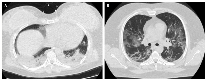
Results: Patients with probable Delta variant had significantly higher CT severity scores, lower PaO2/FiO2 ratio and higher C-reactive protein and lactate dehydrogenase levels at admission. On multivariate analysis, probable Delta variant infection was associated with higher CT severity score. Ground glass opacities and crazy paving patterns were more frequently noticed than consolidation, with the latter being more frequent in Delta cohort, even though not significantly. According to prevalent imaging pattern, the consolidation one was significantly associated with pregnancy (p=0.008).
Conclusions: Patients admitted during predominance of Delta variant circulation had a more severe lung involvement compared to patients in infected when Alpha variant was predominant. Despite imaging pattern seems to be not influenced by viral variant and other clinical variables, the consolidative pattern was observed more frequently in pregnancy.
Keywords: SARS-CoV-2; computed tomography; pneumonia; variant of concern.
Conflict of interest statement
Conflict of interest All authors declare no conflict of interest.
Figures
Similar articles
-
Imaging Severity COVID-19 Assessment in Vaccinated and Unvaccinated Patients: Comparison of the Different Variants in a High Volume Italian Reference Center.J Pers Med. 2022 Jun 10;12(6):955. doi: 10.3390/jpm12060955. J Pers Med. 2022. PMID: 35743740 Free PMC article.
-
Possible Alterations of Imaging Patterns in Computed Tomography for Delta-VOC of SARS-CoV-2.Rofo. 2022 Nov;194(11):1229-1241. doi: 10.1055/a-1826-0436. Epub 2022 Jul 18. Rofo. 2022. PMID: 35850138 English.
-
Comparison of CT findings of coronavirus disease 2019 (COVID-19) pneumonia caused by different major variants.Jpn J Radiol. 2022 Dec;40(12):1246-1256. doi: 10.1007/s11604-022-01301-1. Epub 2022 Jun 28. Jpn J Radiol. 2022. PMID: 35763239 Free PMC article.
-
[Spatial and temporal distribution and predictive value of chest CT scoring in patients with COVID-19].Zhonghua Jie He He Hu Xi Za Zhi. 2021 Mar 12;44(3):230-236. doi: 10.3760/cma.j.cn112147-20200522-00626. Zhonghua Jie He He Hu Xi Za Zhi. 2021. PMID: 33721937 Chinese.
-
Similarities and Differences of Early Pulmonary CT Features of Pneumonia Caused by SARS-CoV-2, SARS-CoV and MERS-CoV: Comparison Based on a Systemic Review.Chin Med Sci J. 2020 Sep 30;35(3):254-261. doi: 10.24920/003727. Chin Med Sci J. 2020. PMID: 32972503 Free PMC article.
Cited by
-
Omicron variant and pulmonary involvements: a chest imaging analysis in asymptomatic and mild COVID-19.Front Public Health. 2024 Jul 4;12:1325474. doi: 10.3389/fpubh.2024.1325474. eCollection 2024. Front Public Health. 2024. PMID: 39035180 Free PMC article.
References
-
- Qi X, Liu Y, Wang J, et al. Clinical course and risk factors for mortality of COVID-19 patients with pre-existing cirrhosis: a multicentre cohort study. Gut [Internet] 2020. May 20, [cited 2020 Jun 26];gutjnl-2020-321666. Available from: https://gut.bmj.com/content/early/2020/06/02/gutjnl-2020-321666. - PMC - PubMed
-
- Nacoti M, Ciocca A, Giupponi A, et al. At the Epicenter of the Covid-19 pandemic and humanitarian crises in Italy: changing perspectives on preparation and mitigation. NEJM Catal. 2020:1–5. Figure 1.
-
- Yang W, Sirajuddin A, Zhang X, et al. The role of imaging in 2019 novel coronavirus pneumonia (COVID-19) Eur Radiol. 2020;30(9):4874–4882. Available from: https://link.springer.com/article/10.1007/s00330-020-06827-4. - DOI - PMC - PubMed
-
- Ye Z, Zhang Y, Wang Y, Huang Z, Song B. Chest CT manifestations of new coronavirus disease 2019 (COVID-19): a pictorial review. Eur Radiol [Internet] 2020 Aug 1;30(8):4381–4389. [cited 2022 Feb 14] Available from: https://link.springer.com/article/10.1007/s00330-020-06801-0. - DOI - PMC - PubMed
LinkOut - more resources
Full Text Sources
Research Materials
Miscellaneous