

This is a preprint.
Non-invasive diagnostic method to objectively measure olfaction and diagnose smell disorders by molecularly targeted fluorescent imaging agent
- PMID: 36482968
- PMCID: PMC9727758
- DOI: 10.1101/2021.10.07.463532
Non-invasive diagnostic method to objectively measure olfaction and diagnose smell disorders by molecularly targeted fluorescent imaging agent
Update in
-
Noninvasive Diagnostic Method to Objectively Measure Olfaction and Diagnose Smell Disorders by a Molecularly Targeted Fluorescence Imaging Agent.J Nucl Med. 2024 Aug 1;65(8):1293-1300. doi: 10.2967/jnumed.123.266123. J Nucl Med. 2024. PMID: 38960711 Free PMC article.
Abstract
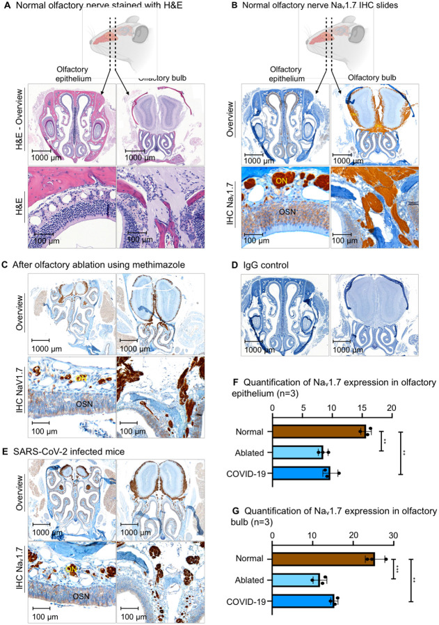
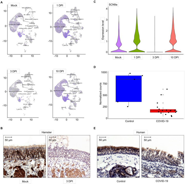
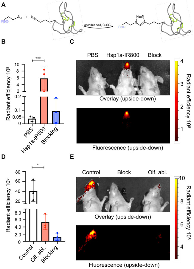
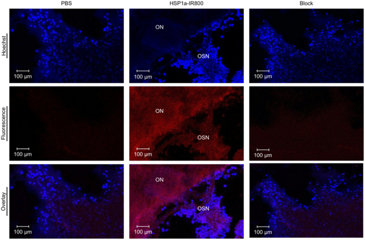
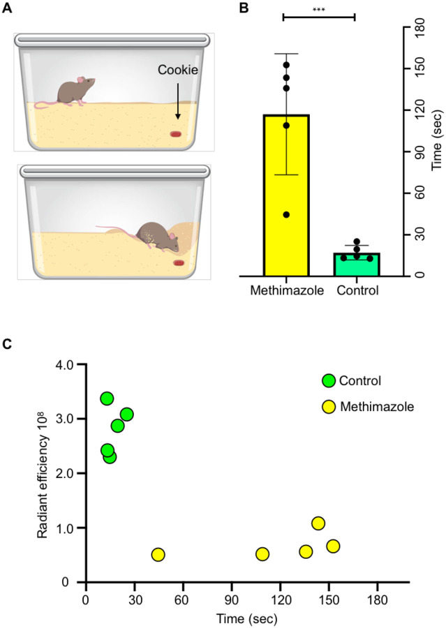
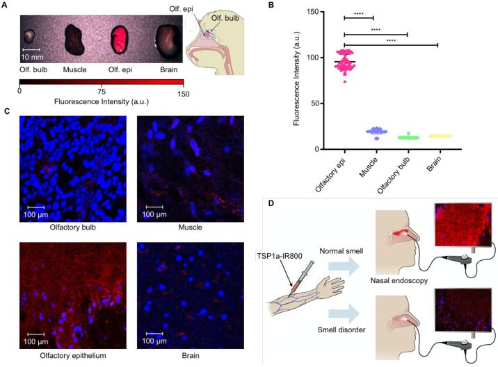
The sense of smell (olfaction) is one of the most important senses for animals including humans. Despite significant advances in the understanding mechanism of olfaction, currently, there are no objective non-invasive methods that can identify loss of smell. Covid-19-related loss of smell has highlighted the need to develop methods that can identify loss of olfaction. Voltage-gated sodium channel 1.7 (NaV1.7) plays a critical role in olfaction by aiding the signal propagation to the olfactory bulb. We have identified several conditions such as chronic inflammation and viral infections such as Covid-19 that lead to loss of smell correlate with downregulation of NaV1.7 expression at transcript and protein levels in the olfactory epithelium. Leveraging this knowledge, we have developed a novel fluorescent probe Tsp1a-IR800 that targets NaV1.7. Using fluorescence imaging we can objectively measure the loss of sense of smell in live animals non-invasively. We also demonstrate that our non-invasive method is semiquantitative because the loss of fluorescence intensity correlates with the level of smell loss. Our results indicate, that our probe Tsp1a-IR800, can objectively diagnose anosmia in animal and human subjects using infrared fluorescence. We believe this method to non-invasively diagnose loss of smell objectively is a significant advancement in relation to current methods that rely on highly subjective behavioral studies and can aid in studying olfaction loss and the development of therapeutic interventions.
Conflict of interest statement
Disclosure of Potential Conflicts of Interest S.P. and T.R. are shareholders of Summit Biomedical Imaging, T.R. is now an executive of and shareholder in Novartis AG. J.G., P.D.S.F., G.K. and T.R. are co-inventors on a Tsp1a-related patent application. All other authors have no conflicts to declare.
Figures
Similar articles
-
Noninvasive Diagnostic Method to Objectively Measure Olfaction and Diagnose Smell Disorders by a Molecularly Targeted Fluorescence Imaging Agent.J Nucl Med. 2024 Aug 1;65(8):1293-1300. doi: 10.2967/jnumed.123.266123. J Nucl Med. 2024. PMID: 38960711 Free PMC article.
-
Persistent post-COVID-19 smell loss is associated with immune cell infiltration and altered gene expression in olfactory epithelium.Sci Transl Med. 2022 Dec 21;14(676):eadd0484. doi: 10.1126/scitranslmed.add0484. Epub 2022 Dec 21. Sci Transl Med. 2022. PMID: 36542694 Free PMC article.
-
Objective Sensory Testing Methods Reveal a Higher Prevalence of Olfactory Loss in COVID-19-Positive Patients Compared to Subjective Methods: A Systematic Review and Meta-Analysis.Chem Senses. 2020 Dec 5;45(9):865-874. doi: 10.1093/chemse/bjaa064. Chem Senses. 2020. PMID: 33245136 Free PMC article.
-
Link between pain and olfaction in an inherited sodium channelopathy.Arch Neurol. 2012 Sep;69(9):1119-23. doi: 10.1001/archneurol.2012.21. Arch Neurol. 2012. PMID: 22733046 Review.
-
Impact of the smell loss on the quality of life and adopted coping strategies in COVID-19 patients.Eur Arch Otorhinolaryngol. 2021 Sep;278(9):3307-3314. doi: 10.1007/s00405-020-06575-7. Epub 2021 Jan 19. Eur Arch Otorhinolaryngol. 2021. PMID: 33464401 Free PMC article.
References
Publication types
Grants and funding
LinkOut - more resources
Full Text Sources