

Early screening model for mild cognitive impairment based on resting-state functional connectivity: a functional near-infrared spectroscopy study
- PMID: 36483024
- PMCID: PMC9722394
- DOI: 10.1117/1.NPh.9.4.045010
Early screening model for mild cognitive impairment based on resting-state functional connectivity: a functional near-infrared spectroscopy study
Abstract
Significance: As an early stage of Alzheimer's disease (AD), the diagnosis of amnestic mild cognitive impairment (aMCI) has important clinical value for timely intervention of AD. Functional near-infrared spectroscopy (fNIRS)-based resting-state brain connectivity analysis, which could provide an economic and quick screening strategy for aMCI, remains to be extensively investigated.
Aim: This study aimed to verify the feasibility of fNIRS-based resting-state brain connectivity for evaluating brain function in patients with aMCI, and to determine an early screening model for auxiliary diagnosis.
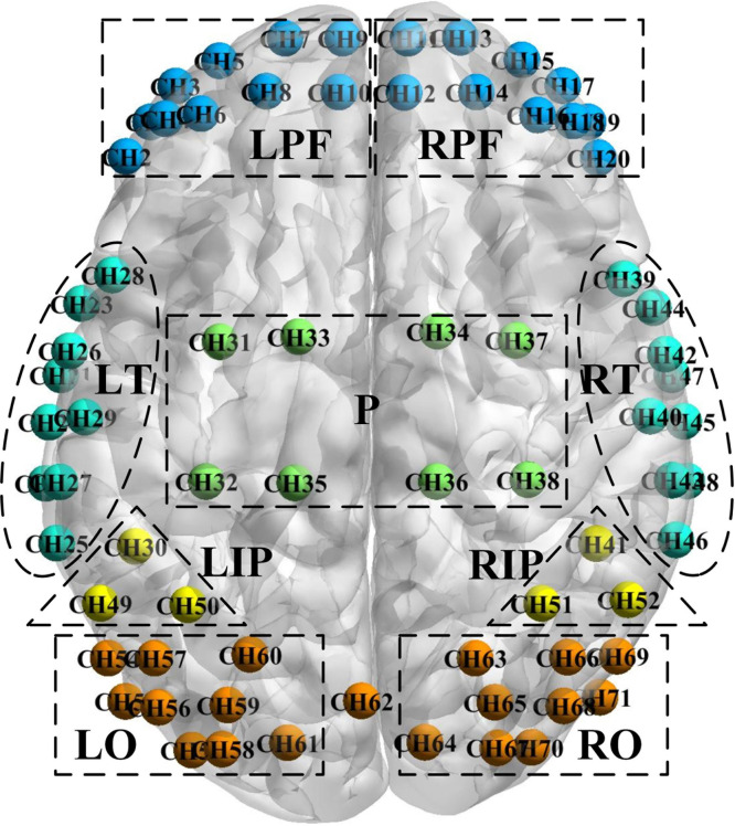
Approach: The resting-state fNIRS was utilized for exploring the changes in functional connectivity of 64 patients with aMCI. The region of interest (ROI)-based and channel-based connections with significant inter-group differences have been extracted through the two-sample -tests and the receiver operating characteristic (ROC). These connections with specificity and sensitivity were then taken as features for classification.
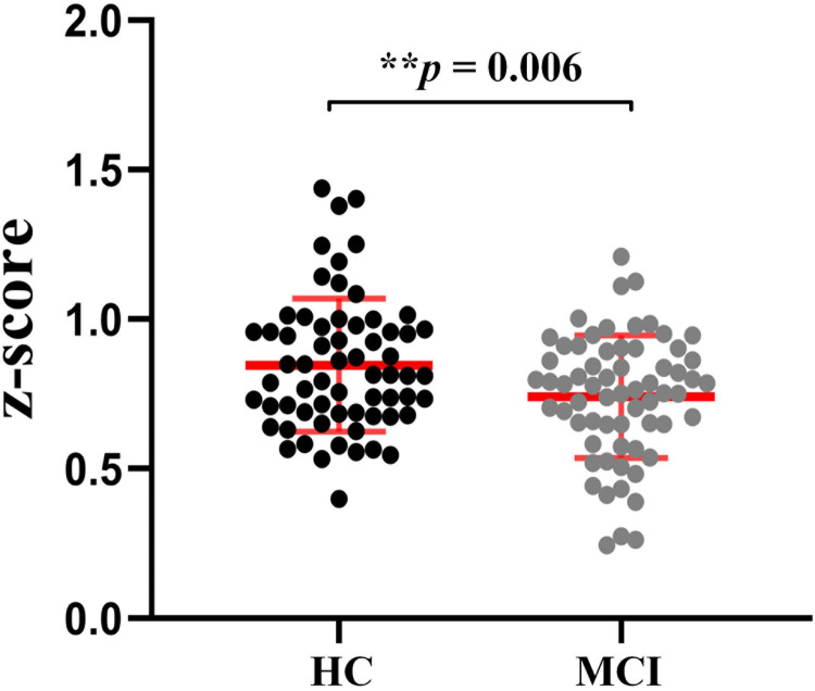
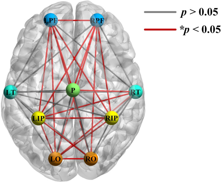
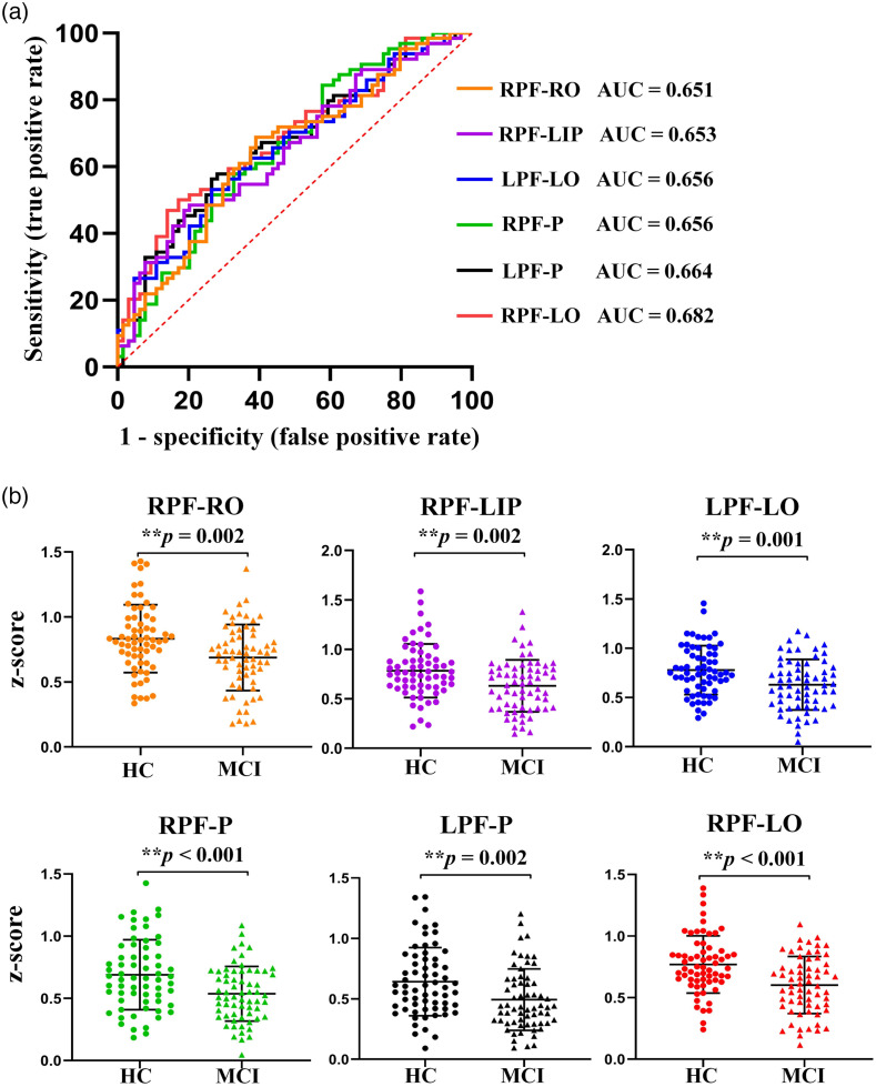
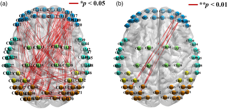
Results: Compared with healthy controls, connections of the MCI group were significantly reduced between the bilateral prefrontal, parietal, occipital, and right temporal lobes. Specifically, the long-range connections from prefrontal to occipital lobe, and from prefrontal to parietal lobe, exhibited stronger identifiability (area under the ROC curve , ** ). Subsequently, the optimal classification accuracy of ROI-based connections was 71.59%. Furthermore, the most responsive connections were located between the right dorsolateral prefrontal lobe and the left occipital lobe, concomitant with the highest classification accuracy of 73.86%.
Conclusion: Our findings indicate that fNIRS-based resting-state functional connectivity analysis could support MCI diagnosis. Notably, long-range connections involving the prefrontal and occipital lobes have the potential to be efficient biomarkers.
Keywords: amnesic mild cognitive impairment; functional connectivity; functional near-infrared spectroscopy; resting state.
© 2022 The Authors.
Figures
Similar articles
-
An exploration of distinguishing subjective cognitive decline and mild cognitive impairment based on resting-state prefrontal functional connectivity assessed by functional near-infrared spectroscopy.Front Aging Neurosci. 2025 Jan 8;16:1468246. doi: 10.3389/fnagi.2024.1468246. eCollection 2024. Front Aging Neurosci. 2025. PMID: 39845444 Free PMC article.
-
Correlation Between Prefrontal Functional Connectivity and the Degree of Cognitive Impairment in Alzheimer's Disease: A Functional Near-Infrared Spectroscopy Study.J Alzheimers Dis. 2024;98(4):1287-1300. doi: 10.3233/JAD-230648. J Alzheimers Dis. 2024. PMID: 38517784
-
Comparative study of brain functional imaging of brain in patients with mild to moderate Alzheimer's disease based on functional near infrared spectroscopy.BMC Neurol. 2025 Apr 28;25(1):186. doi: 10.1186/s12883-024-03989-2. BMC Neurol. 2025. PMID: 40289104 Free PMC article.
-
Identification of functional near-infrared spectroscopy for older adults with mild cognitive impairment: a systematic review.Front Aging Neurosci. 2025 Apr 9;17:1492800. doi: 10.3389/fnagi.2025.1492800. eCollection 2025. Front Aging Neurosci. 2025. PMID: 40271185 Free PMC article.
-
Resting-state functional near-infrared spectroscopy in neurodegenerative diseases - A systematic review.Neuroimage Clin. 2025;45:103733. doi: 10.1016/j.nicl.2025.103733. Epub 2025 Jan 17. Neuroimage Clin. 2025. PMID: 39889542 Free PMC article.
Cited by
-
Alterations in brain functional connectivity in patients with mild cognitive impairment: A systematic review and meta-analysis of functional near-infrared spectroscopy studies.Brain Behav. 2024 Apr;14(4):e3414. doi: 10.1002/brb3.3414. Brain Behav. 2024. PMID: 38616330 Free PMC article.
-
Alterations in brain function in patients with post-stroke cognitive impairment: a resting-state functional magnetic resonance imaging study.Front Aging Neurosci. 2025 Feb 19;17:1501082. doi: 10.3389/fnagi.2025.1501082. eCollection 2025. Front Aging Neurosci. 2025. PMID: 40046780 Free PMC article.
-
The analysis of brain functional connectivity of post-stroke cognitive impairment patients: an fNIRS study.Front Neurosci. 2023 May 5;17:1168773. doi: 10.3389/fnins.2023.1168773. eCollection 2023. Front Neurosci. 2023. PMID: 37214384 Free PMC article.
-
An exploration of distinguishing subjective cognitive decline and mild cognitive impairment based on resting-state prefrontal functional connectivity assessed by functional near-infrared spectroscopy.Front Aging Neurosci. 2025 Jan 8;16:1468246. doi: 10.3389/fnagi.2024.1468246. eCollection 2024. Front Aging Neurosci. 2025. PMID: 39845444 Free PMC article.
-
Functional near-infrared spectroscopy in elderly patients with four types of dementia.World J Psychiatry. 2023 May 19;13(5):203-214. doi: 10.5498/wjp.v13.i5.203. eCollection 2023 May 19. World J Psychiatry. 2023. PMID: 37303929 Free PMC article.
References
-
- Patterson C., World Alzheimer Report 2018-The State of the Art of Dementia Research: New Frontiers, Alzheimer’s Disease International (ADI), London, UK: (2018).
LinkOut - more resources
Full Text Sources
Research Materials
