

MIR4435-2HG in exosomes promotes gastric carcinogenesis by inducing M2 polarization in macrophages
- PMID: 36483041
- PMCID: PMC9723220
- DOI: 10.3389/fonc.2022.1017745
MIR4435-2HG in exosomes promotes gastric carcinogenesis by inducing M2 polarization in macrophages
Abstract
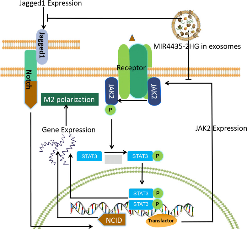
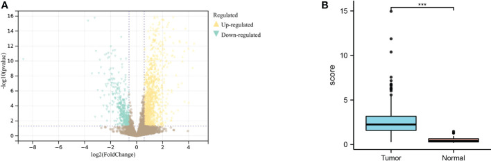
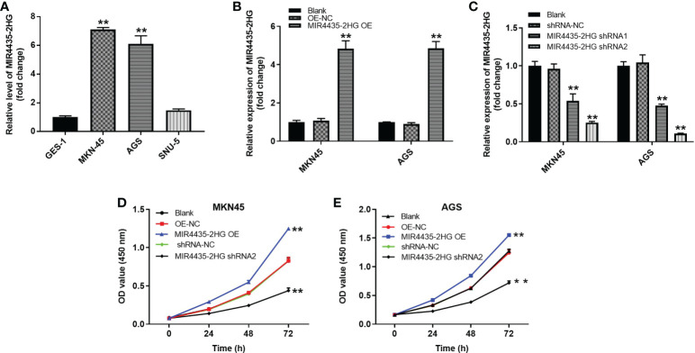
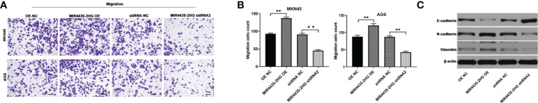
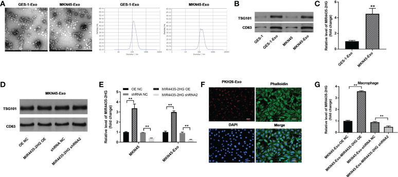
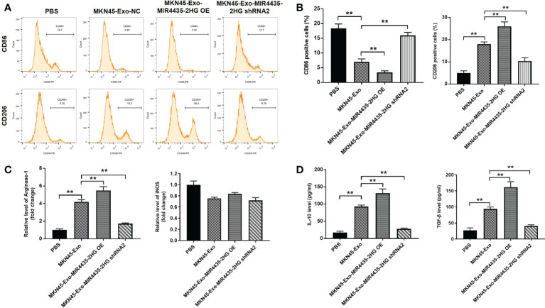
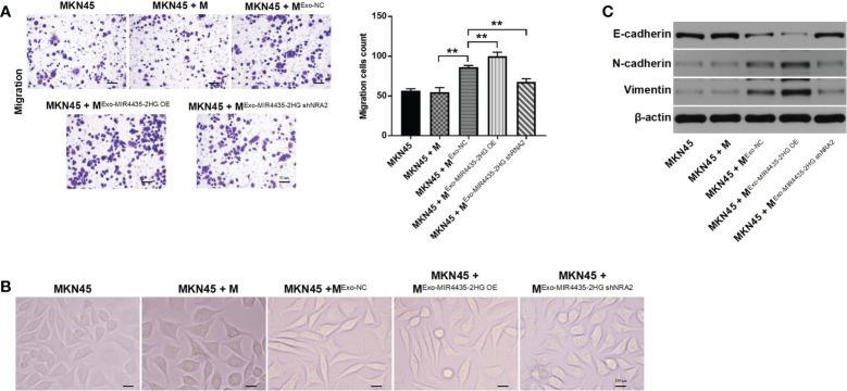
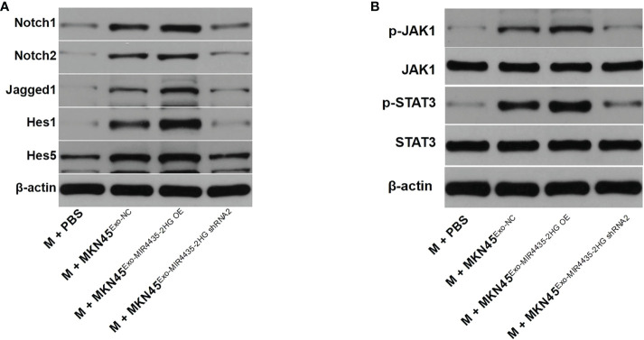
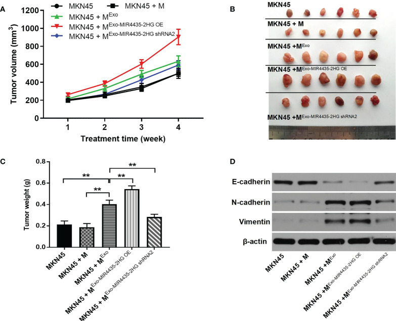
Gastric cancer (GC) is a cancer with a high mortality rate. lncRNAs play a role in regulating GC tumorigenesis. In this paper, we analyzed differentially expressed lncRNAs between GC and adjacent normal tissues using multiple bioinformatics tools to identify new potential targets in GC. Cell viability and migration ability were detected using the Cell Counting Kit-8 (CCK-8) and transwell assays, MIR4435-2HG was negatively correlated with the survival rate of GC patients, and by inhibiting the activity of MIR4435-2HG, the viability and migration ability of GC cells could be reduced. In addition, RT- qPCR and western blot to detect gene and protein level expression, transmission electron microscopy and nanoparticle tracking analysis (NTA) to study the efficiency of exosome isolation, and flow cytometry to observe cell differentiation were employed, delivery of MIR4435-2HG shRNA via MKN45 cell-derived exosomes significantly reversed the MKN45 exosome-induced M2 polarization in macrophages. Furthermore, the low expression of MIR4435-2HG in MKN45 cell-derived exosomes inhibited the Jagged1/Notch and JAK1/STAT3 pathways in macrophages; MIR4435-2HG downregulated exosomes were found to significantly inhibit GC tumor growth in vivo by establishing a mouse model. In short, MKN45 cell-derived exosomes deliver lncRNA MIR4435-2HG, which promotes gastric carcinogenesis by inducing macrophage M2 polarization.
Keywords: GC; JAK1/STAT3; Jagged1/Notch; MIR4435-2HG; MKN45.
Copyright © 2022 Li, Chen, Gao, Tang, Zhou, Zhang, Zhang, Shen, Guo and Fu.
Conflict of interest statement
The authors declare that the research was conducted in the absence of any commercial or financial relationships that could be construed as a potential conflict of interest.
Figures
Similar articles
-
M2 Macrophage-Derived Exosomal lncRNA MIR4435-2HG Promotes Progression of Infantile Hemangiomas by Targeting HNRNPA1.Int J Nanomedicine. 2023 Oct 20;18:5943-5960. doi: 10.2147/IJN.S435132. eCollection 2023. Int J Nanomedicine. 2023. PMID: 37881607 Free PMC article.
-
Inhibition of MIR4435-2HG on Invasion, Migration, and EMT of Gastric Carcinoma Cells by Mediating MiR-138-5p/Sox4 Axis.Front Oncol. 2021 Aug 31;11:661288. doi: 10.3389/fonc.2021.661288. eCollection 2021. Front Oncol. 2021. PMID: 34532282 Free PMC article.
-
LncRNA MIR4435-2HG promotes proliferation, migration, invasion and epithelial mesenchymal transition via targeting miR-22-3p/TMEM9B in breast cancer.Am J Transl Res. 2022 Aug 15;14(8):5441-5454. eCollection 2022. Am J Transl Res. 2022. PMID: 36105009 Free PMC article.
-
MIR4435-2HG: A Tumor-associated Long Non-coding RNA.Curr Pharm Des. 2022;28(25):2043-2051. doi: 10.2174/1381612828666220607100228. Curr Pharm Des. 2022. PMID: 35674305 Review.
-
MIR4435-2HG: A newly proposed lncRNA in human cancer.Biomed Pharmacother. 2022 Jun;150:112971. doi: 10.1016/j.biopha.2022.112971. Epub 2022 Apr 18. Biomed Pharmacother. 2022. PMID: 35447550 Review.
Cited by
-
The Emerging Role of LncRNA AWPPH in Multiple Cancers: A Review Study.Curr Mol Med. 2025;25(3):237-268. doi: 10.2174/1566524023666230816163031. Curr Mol Med. 2025. PMID: 37587826 Review.
-
Biological Roles and Pathogenic Mechanisms of LncRNA MIR4435-2HG in Cancer: A Comprehensive Review.Curr Issues Mol Biol. 2023 Nov 4;45(11):8864-8881. doi: 10.3390/cimb45110556. Curr Issues Mol Biol. 2023. PMID: 37998733 Free PMC article. Review.
-
Tumour-associated macrophages in gastric cancer: From function and mechanism to application.Clin Transl Med. 2023 Aug;13(8):e1386. doi: 10.1002/ctm2.1386. Clin Transl Med. 2023. PMID: 37608500 Free PMC article. Review.
-
N‑methyladenosine reader YTHDF2‑mediated AC026691.1 degradation promotes gastric cancer cell proliferation, migration and M2 macrophage polarization.Mol Med Rep. 2025 May;31(5):120. doi: 10.3892/mmr.2025.13485. Epub 2025 Mar 7. Mol Med Rep. 2025. PMID: 40052573 Free PMC article.
-
Exosomal LncRNAs in Gastrointestinal Cancer: Biological Functions and Emerging Clinical Applications.Cancers (Basel). 2023 Feb 2;15(3):959. doi: 10.3390/cancers15030959. Cancers (Basel). 2023. PMID: 36765913 Free PMC article. Review.
References
LinkOut - more resources
Full Text Sources
Research Materials
Miscellaneous