

The mechanisms underlying the immune control of Zika virus infection at the maternal-fetal interface
- PMID: 36483552
- PMCID: PMC9723234
- DOI: 10.3389/fimmu.2022.1000861
The mechanisms underlying the immune control of Zika virus infection at the maternal-fetal interface
Abstract
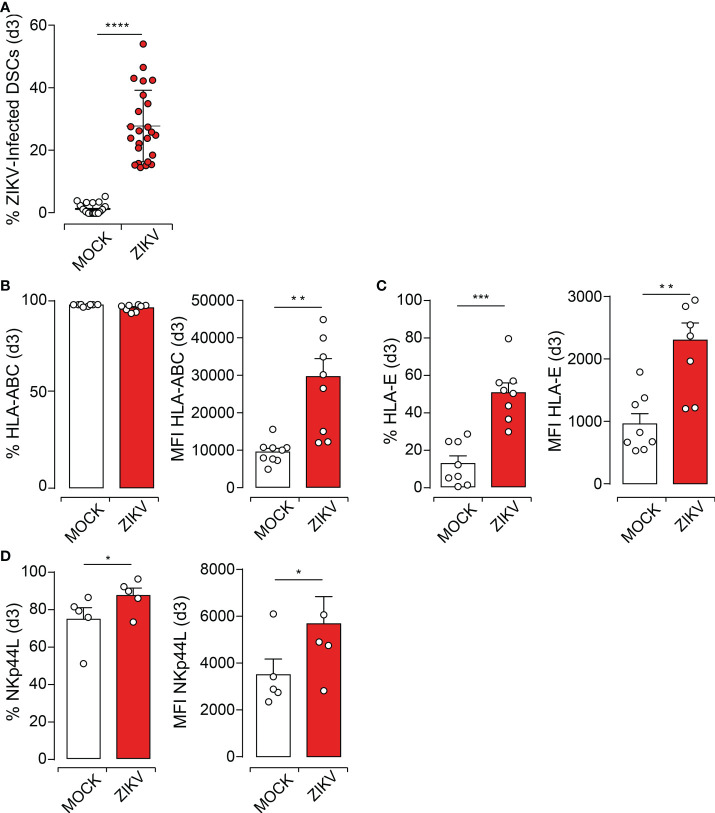
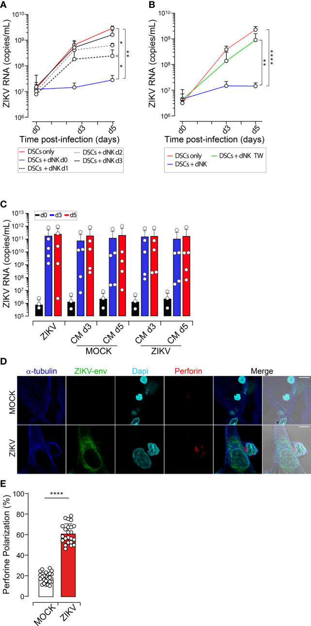
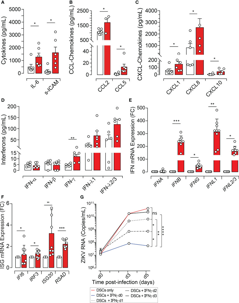
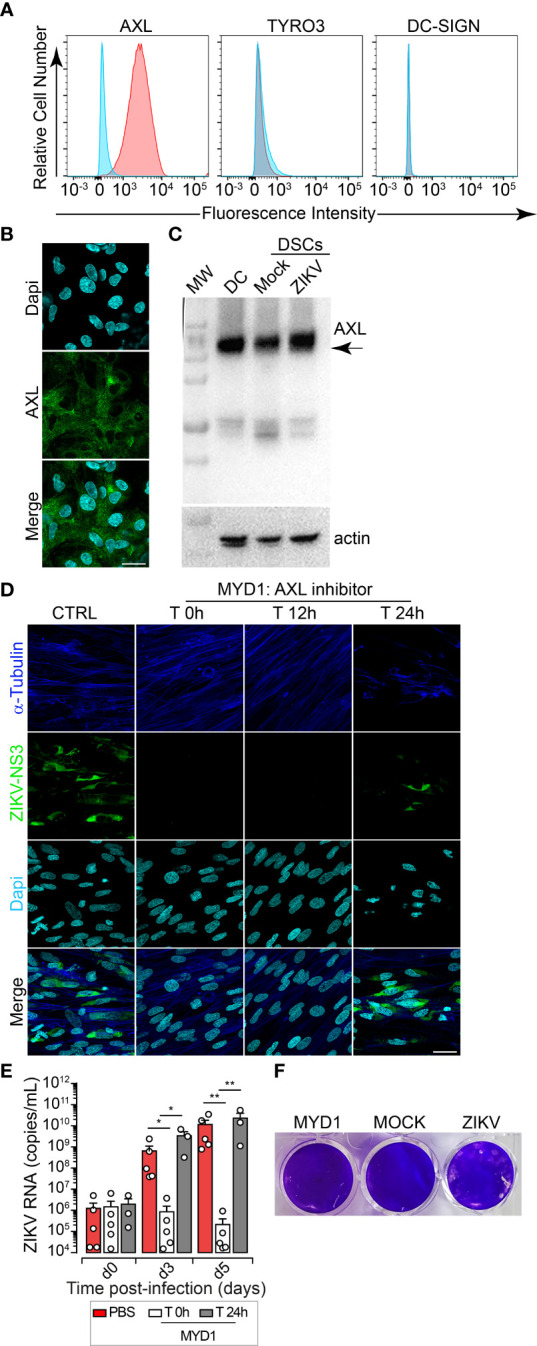
Unlike other Flaviviruses, Zika virus (ZIKV) infection during the first trimester of pregnancy causes severe pregnancy outcomes including the devastating microcephaly and diseases associated with placental dysfunctions. We have previously reported that the maternal decidua basalis, the major maternal-fetal interface, serves as a replication platform enabling virus amplification before dissemination to the fetal compartment. However, the rate of congenital infection is quite low, suggesting the presence of a natural barrier against viral infection. Using primary cells from first-trimester pregnancy samples, we investigated in this study how the maternal decidua can interfere with ZIKV infection. Our study reveals that whether through their interactions with dNK cells, the main immune cell population of the first-trimester decidua, or their production of proinflammatory cytokines, decidual stromal cells (DSCs) are the main regulators of ZIKV infection during pregnancy. We also validate the functional role of AXL as a crucial receptor for ZIKV entry in DSCs and demonstrate that targeted inhibition of ligand-receptor interaction at the early stage of the infection is effective in drastically reducing virus pathogenesis at the maternal-fetal interface. Collectively, our results provide insights into the mechanisms through which ZIKV infection and spreading can be limited. The strategy of circumventing viral entry at the maternal-fetus interface limits virus dissemination to fetal tissues, thereby preventing congenital abnormalities.
Keywords: Zika virus; infection; inflammation; maternal-fetal interface; natural killer cells.
Copyright © 2022 Espino, Gouilly, Chen, Colin, Guerby, Izopet, Amara, Tabiasco, Al-Daccak, El Costa and Jabrane-Ferrat.
Conflict of interest statement
The authors declare that the research was conducted in the absence of any commercial or financial relationships that could be construed as a potential conflict of interest.
Figures
Similar articles
-
Zika Virus Infects Early- and Midgestation Human Maternal Decidual Tissues, Inducing Distinct Innate Tissue Responses in the Maternal-Fetal Interface.J Virol. 2017 Jan 31;91(4):e01905-16. doi: 10.1128/JVI.01905-16. Print 2017 Feb 15. J Virol. 2017. PMID: 27974560 Free PMC article.
-
Zika Virus Targets Multiple Tissues and Cell Types During the First Trimester of Pregnancy.Methods Mol Biol. 2020;2142:235-249. doi: 10.1007/978-1-0716-0581-3_18. Methods Mol Biol. 2020. PMID: 32367371
-
Immune Profile of the Normal Maternal-Fetal Interface in Rhesus Macaques and Its Alteration Following Zika Virus Infection.Front Immunol. 2021 Jul 29;12:719810. doi: 10.3389/fimmu.2021.719810. eCollection 2021. Front Immunol. 2021. PMID: 34394129 Free PMC article.
-
Immune Evasion Strategies Used by Zika Virus to Infect the Fetal Eye and Brain.Viral Immunol. 2020 Jan/Feb;33(1):22-37. doi: 10.1089/vim.2019.0082. Epub 2019 Nov 5. Viral Immunol. 2020. PMID: 31687902 Free PMC article. Review.
-
Maternal-Fetal Interplay in Zika Virus Infection and Adverse Perinatal Outcomes.Front Immunol. 2020 Feb 14;11:175. doi: 10.3389/fimmu.2020.00175. eCollection 2020. Front Immunol. 2020. PMID: 32117303 Free PMC article. Review.
Cited by
-
Coinfection with chikungunya and Zika results in mild disease and distinct inflammatory response.Npj Viruses. 2025 Feb 11;3(1):10. doi: 10.1038/s44298-025-00098-w. Npj Viruses. 2025. PMID: 40295813 Free PMC article.
-
The Evolving Role of Zika Virus Envelope Protein in Viral Entry and Pathogenesis.Viruses. 2025 Jun 6;17(6):817. doi: 10.3390/v17060817. Viruses. 2025. PMID: 40573410 Free PMC article. Review.
-
Fetal Zika virus inoculation in macaques revealed control of the fetal viral load during pregnancy.Virol J. 2024 Sep 3;21(1):209. doi: 10.1186/s12985-024-02468-x. Virol J. 2024. PMID: 39227837 Free PMC article.
-
Insight into the Natural Biomolecules (BMs): Promising Candidates as Zika Virus Inhibitors.Infect Disord Drug Targets. 2024;24(7):e020224226681. doi: 10.2174/0118715265272414231226092146. Infect Disord Drug Targets. 2024. PMID: 38318833 Review.
-
Congenital Zika virus infection in laboratory animals: a comparative review highlights translational studies on the maternal-foetal interface.Mem Inst Oswaldo Cruz. 2025 Feb 28;120:e240125. doi: 10.1590/0074-02760240125. eCollection 2025. Mem Inst Oswaldo Cruz. 2025. PMID: 40052994 Free PMC article. Review.
References
-
- El Costa H, Tabiasco J, Berrebi A, Parant O, Aguerre-Girr M, Piccinni MP, et al. . Effector functions of human decidual NK cells in healthy early pregnancy are dependent on the specific engagement of natural cytotoxicity receptors. J Reprod Immunol (2009) 82(2):142–7. doi: 10.1016/j.jri.2009.06.123 - DOI - PubMed
Publication types
MeSH terms
LinkOut - more resources
Full Text Sources
Medical
Research Materials
Miscellaneous