Review
. 2022 Nov;11(6):564-582.
doi: 10.21037/acs-2022-rmvs-26.

Historical evolution of robot-assisted cardiac surgery: a 25-year journey

Affiliations
Review

Historical evolution of robot-assisted cardiac surgery: a 25-year journey

W Randolph Chitwood Jr. Ann Cardiothorac Surg. 2022 Nov.

Abstract

Many patients and surgeons today favor the least invasive access to an operative site. The adoption of robot-assisted cardiac surgery has been slow, but now has come to fruition. The development of modern surgical robots took surgeons close collaboration with mechanical, electrical, and optical engineers. Moreover, the necessary project funding required entrepreneurs, federal grants, and venture capital. Non-robotic minimally invasive cardiac surgery paved the way to the application of surgical robots by making changes in operative approaches, instruments, visioning modalities, cardiopulmonary perfusion techniques, and especially surgeons' attitudes. In this article, the serial development of robot-assisted cardiac surgery is detailed from the beginning and through clinical application. Included are references to the historical and most recent clinical series that have given us the evidence that robot-assisted cardiac surgery is safe and provides excellent outcomes. To this end, in many institutions these procedures now have become a new standard of care. This evolution reflects Sir Isaac Newton's famous 1676 quote when referring to Rene Descartes, "If have seen further [sic] than others, it is by standing on the shoulders of giants".

Keywords: Cardiac; history; minimally invasive; robotic.


Conflict of interest statement

Conflicts of Interest: The author declares no conflicts of interest.

Figures
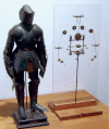
Figure 1
Figure 1
Leonardo daVinci’s 1495 mechanical knight. Note that the activating pulley and cable mechanisms were very close to those in the modern daVinci Surgical System.
Figure 2
Figure 2
1995 original US Patent 5725496 by William Peters MD (Stanford University) —endoballoon aortic occlusion catheter. This concept was adopted for the commercial Heartport Port Access System.
Figure 3
Figure 3
Development of endoballoon aortic occlusion. (A) The first Port Access mitral valve replacement. (B) The Stanford University and Malaysia team that performed the first four Heartport Port Access mitral valve replacements in October 1996. (C) The original Heartport System that consisted of an aortic occluder endoballoon, a retrograde coronary sinus cardioplegia catheter, and a pulmonary artery vent. The retrograde femoral artery perfusion cannula was designed with a side-arm valve to introduce the balloon catheter.
Figure 4
Figure 4
The author is wearing the Vista head-mounted display for 3-D vision during an early minimally invasive mitral repair.
Figure 5
Figure 5
First video-assisted minimally invasive mitral valve replacement in the United States. (A) Mini-thoracotomy with venous cannula placed through the incision—original trans-thoracic cross clamp—5 mm endoscope. (B) Operating using 2-dimensional endoscopic vision. (C) St. Jude mechanical prosthesis—sewing ring sutures were tied with the “wire” from a Rummel tourniquet. Thereafter, new instruments were designed for this purpose.
Figure 6
Figure 6
The first surgical robot prototype. (A) Green telepresence surgeon’s workstation with hand controls—Stanford Research Institute 1994. (B) Exchangeable end-effector surgical instruments. The figure is reproduced with permission from the Journal of Laparoscopic Surgery and Society of Laparoscopic and Robotic Surgeon.
Figure 7
Figure 7
The author performing a video-assisted minimally invasive mitral valve repair using the AESOP 3000 to voice control the endoscope position.
Figure 8
Figure 8
The ZEUS operating console with voice activated endoscope position control.
Figure 9
Figure 9
Intuitive surgical instrument arm development. (A) Lenny the first protype; (B) Mona—the first prototype used for clinical studies; (C) the first daVinci commercial arm; (D) daVinci S and Si commercial arms.
Figure 10
Figure 10
Mona-daVinci prototype. (A) The author operating at the daVinci prototype (Mona) console—in 1997. (B) Early wristed robot instruments with Mona.
Figure 11
Figure 11
The team that performed the first robot-assisted mitral valve repair at Broussais Hôpital Paris—May 7, 1998. Second row L to R: Professor Carpentier, Dr. Fred Moll (Founder of Intuitive Surgical, Inc.). Third row L to R: D. Didier Loulmet, Dr. Albert Starr.
Figure 12
Figure 12
The first of five robot-assisted mitral valve repairs done at the Leipzig Heart Center—May 27, 1998. (A) Professor Fredrich Mohr operating from the prototype surgeon console—Mona. (B) Surgical Team L to R—Frau Ingrid Conradt (Chief Scrub Nurse), Dave Rosa (Intuitive Engineer), Volkmar Falk (Surgeon)—Mona Instrument Cart at operating table during an instrument exchange. (C) An annuloplasty ring being implanted. Note the small inset shows simultaneous Professor Mohr’s hand motions.
Figure 13
Figure 13
The first commercial daVinci Surgical System in the United States. (A) Dr. Wiley Nifong has just received the new daVinci robot. (B) The first daVinci robotic research and training laboratory 1999—East Carolina University School of Medicine. (C) Beginning to develop our method for performing daVinci-assisted mitral valve surgery. Dr. Wiley Nifong at the instrument cart and Dr. Joseph Elberry at the surgeon console.
Figure 14
Figure 14
First daVinci Robot mitral repair in North America. (A) The operating team during the operation. (B) A quadrangular posterior leaflet resection which was followed by implantation of an annuloplasty band. (C) Our first patient three days post mitral repair. She was enrolled in the first FDA safety and efficacy trial. She is alive and well 22 years later at the age of 92. Informed consent was obtained from the patient to publish this picture. FDA, Food and Drug Administration.
Figure 15
Figure 15
daVinci XI operating room at the Plano/Baylor Heart Hospital—Dr. Robert Smith repairing a mitral valve.
Figure 16
Figure 16
daVinci XI instrument arms. (A) Multiple instrument arm positions. (B) Instrument arms are docked at the operating table with instruments inserted for a mitral valve repair. Note the small working port for the assistant.
Video
Video
Robotic Mitral Surgery - Lessons Learnt.

References

    1. Čapek K. Rossumovi Univerzální Roboti (Rossum’s Universal Robots). Prague, 1921.
    1. Kolessov VI. Mammary artery-coronary artery anastomosis as method of treatment for angina pectoris. J Thorac Cardiovasc Surg 1967;54:535-44. 10.1016/S0022-5223(19)43061-4 - DOI - PubMed
    1. Ankeny JL. Editorial: To use or not to use the pump oxygenator in coronary bypass operations. Ann Thorac Surg 1975;19:108-9. 10.1016/S0003-4975(10)65744-X - DOI - PubMed
    1. Ankeney JL, Goldstein DJ. Off-pump bypass of the left anterior descending coronary artery: 23- to 34-year follow-up. J Thorac Cardiovasc Surg 2007;133:1499-503. 10.1016/j.jtcvs.2007.01.071 - DOI - PubMed
    1. Buffolo E, de Andrade CS, Branco JN, et al. Coronary artery bypass grafting without cardiopulmonary bypass. Ann Thorac Surg 1996;61:63-6. 10.1016/0003-4975(95)00840-3 - DOI - PubMed