Case discussions of missed traumatic fractures on computed tomography scans
- PMID: 36483672
- PMCID: PMC9724140
- DOI: 10.4102/sajr.v26i1.2516
Case discussions of missed traumatic fractures on computed tomography scans
Abstract
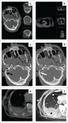
Radiological diagnostic errors are common and may have severe consequences. Understanding these errors and their possible causes is crucial for optimising patient care and improving radiological training. Recent postmortem studies using an animal model highlighted the difficulties associated with accurate fracture diagnosis using radiological imaging. The present study aimed to highlight the fact that certain fractures are easily missed on CT scans in a clinical setting and that caution is advised. A few such cases were discussed to raise the level of suspicion to prevent similar diagnostic errors in future cases. Records of adult patients from the radiological department at an academic hospital in South Africa were retrospectively reviewed. Case studies were selected by identifying records of patients between January and June 2021 where traumatic fractures were missed during initial imaging interpretation but later detected during secondary analysis or on follow-up scans. Seven cases were identified, and the possible causes of the diagnostic errors were evaluated by reviewing the history of each case, level of experience of each reporting radiologist, scan quality and time of day that initial imaging interpretation of each scan was performed. The causes were multifactorial, potentially including a lack of experience, fatigue, heavy workloads or inadequate training of the initial reporting radiologist. Identifying these causes, openly discussing them and providing additional training for radiologists may aid in reducing these errors.
Contribution: This article aimed to use case examples of missed injuries on CT scanning of patients in a South African emergency trauma setting in order to highlight and provide insight into common errors in scan interpretation, their causes and possible means of mitigating them.
Keywords: diagnostic errors; emergency department; fracture misdiagnoses; radiology; traumatic fractures.
© 2022. The Authors.
Conflict of interest statement
The authors declare that they have no financial or personal relationships that may have inappropriately influenced them in writing this article.
Figures


Similar articles
-
[Upper cervical spine fracture: sources of misdiagnosis].Radiol Med. 1999 Oct;98(4):230-5. Radiol Med. 1999. PMID: 10615359 Italian.
-
An audit of the polytrauma fracture detection rate of clinicians evaluating lodox statscan bodygrams in two South African public sector trauma units.Injury. 2019 Sep;50(9):1511-1515. doi: 10.1016/j.injury.2019.07.036. Epub 2019 Jul 30. Injury. 2019. PMID: 31399208
-
Errors in imaging patients in the emergency setting.Br J Radiol. 2016;89(1061):20150914. doi: 10.1259/bjr.20150914. Epub 2016 Feb 3. Br J Radiol. 2016. PMID: 26838955 Free PMC article. Review.
-
Interpretation of emergency CT scans in polytrauma: trauma surgeon vs radiologist.Afr J Emerg Med. 2020 Jun;10(2):90-94. doi: 10.1016/j.afjem.2020.01.008. Epub 2020 Mar 7. Afr J Emerg Med. 2020. PMID: 32612915 Free PMC article.
-
Easily missed pathologies of the musculoskeletal system in the emergency radiology setting.Rofo. 2025 Mar;197(3):277-287. doi: 10.1055/a-2369-8330. Epub 2024 Aug 2. Rofo. 2025. PMID: 39094774 Review. English.
Cited by
-
Automated deep learning-assisted early detection of radiation-induced temporal lobe injury on MRI: a multicenter retrospective analysis.Eur Radiol. 2025 Sep;35(9):5239-5251. doi: 10.1007/s00330-025-11470-y. Epub 2025 Mar 7. Eur Radiol. 2025. PMID: 40050455
References
-
- Clark C, Mole CG, Heyns M. Patterns of blunt force homicide in the West Metropole of the City of Cape Town, South Africa. S Afr J Sci. 2017;113(5–6):1–6. 10.17159/sajs.2017/20160214 - DOI
Publication types
LinkOut - more resources
Full Text Sources
