. 2022 Nov 22:10:926283.
doi: 10.3389/fcell.2022.926283. eCollection 2022.

ALD-R491 regulates vimentin filament stability and solubility, cell contractile force, cell migration speed and directionality

Affiliations

ALD-R491 regulates vimentin filament stability and solubility, cell contractile force, cell migration speed and directionality

Hyejeong Rosemary Kim et al. Front Cell Dev Biol. .

Abstract

Metastasizing cells express the intermediate filament protein vimentin, which is used to diagnose invasive tumors in the clinic. However, the role of vimentin in cell motility, and if the assembly of non-filamentous variants of vimentin into filaments regulates cell migration remains unclear. We observed that the vimentin-targeting drug ALD-R491 increased the stability of vimentin filaments, by reducing filament assembly and/or disassembly. ALD-R491-treatment also resulted in more bundled and disorganized filaments and an increased pool of non-filamentous vimentin. This was accompanied by a reduction in size of cell-matrix adhesions and increased cellular contractile forces. Moreover, during cell migration, cells showed erratic formation of lamellipodia at the cell periphery, loss of coordinated cell movement, reduced cell migration speed, directionality and an elongated cell shape with long thin extensions at the rear that often detached. Taken together, these results indicate that the stability of vimentin filaments and the soluble pool of vimentin regulate the speed and directionality of cell migration and the capacity of cells to migrate in a mechanically cohesive manner. These observations suggest that the stability of vimentin filaments governs the adhesive, physical and migratory properties of cells, and expands our understanding of vimentin functions in health and disease, including cancer metastasis.

Keywords: cell migration; contractile forces; cytoskeletal organization; fluorescence recovery after photobleaching (FRAP); intermediate filaments (IFs); traction force microscopy (TFM); vimentin; vimentin assembly.


Conflict of interest statement

Authors DS and RC were employed by Aluda Pharmaceuticals, Inc. The remaining authors declare that the research was conducted in the absence of any commercial or financial relationships that could be construed as a potential conflict of interest.

Figures

FIGURE 1
FIGURE 1
R491-treatment changes the spatial organization of vimentin filaments. (A) Representative images of BJ-Ras-cells with 5 μM control (top) or R491 (lower panel), showing vimentin, microtubule and actin, as indicated, and merged images of vimentin (red), F-actin (green), tubulin (white) and nuclei (blue). Inset images: magnified views of boxed areas in corresponding images. Scale bars: 10 μm. The graphs show the quantification of (B) the loop circumferences of vimentin filaments in DMSO (n = 23) and R491-treated (n = 20) cells, and (C) the diameter of filaments in DMSO (n = 15) and R491-treated (n = 15) cells. ****p ≤ 0.0001. AU: arbitrary unit. Data presented as mean ± SEM.
FIGURE 2
FIGURE 2
R491-treatment stabilizes vimentin in BJ-Ras cells. (A) Representative FRAP images of cells treated with 5 µM control or R491 3 h prior to imaging. Inset images show magnified views of the bleached areas in the larger images. The FRAP-bleached regions are shown in white circles over time. The blue circle in the top panel indicates a FRAP-bleached region near the cell periphery, in which vimentin filaments moved and therefore were difficult to track (dotted blue circles), and excluded from the data set. (B) FRAP recovery curves of vimentin-EGFP in cells treated with 5 μM control (light blue, n = 14) or R491 (dark blue, n = 13), showing the means of at least 3 biological repeats. One-phase exponential recovery curve was fitted (solid line), and 95% confidence intervals are shown (dotted lines). ****p ≤ 0.0001 drug treated compared with control plateau (Ymax). p = 0.319 for drug-treated as compared with control Rate of recovery (K). Scale bar: 5 µm.
FIGURE 3
FIGURE 3
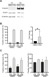
R491-treatment increases the soluble fraction of vimentin in BJ-Ras cells. (A) Representative western blot images of cells with DMSO control- or R491-treatment, analyzed for vimentin, actin and microtubules in soluble (CS) and insoluble (CM) cytoskeletal fractions, as quantified in (B,C). The western blot images are taken from the same membrane. Graphs show means of at least 3 biological repeats. Data presented as mean ± SEM. *p < 0.05.
FIGURE 4
FIGURE 4
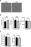
R491-treatment decreases the speed and directionality of cell migration and increase cell elongation in BJ-Ras cells. (A) Oblique-contrast images of BJ-Ras-cells treated with control or R491, as indicated, and quantified with regard to (B) aspect ratio, circularity, spreading area or (C) migration speed and persistence, as indicated. Graphs show means of at least 3 biological repeats. Data presented as mean ± SD. *p ≤ 0.05, **p ≤ 0.01, ***p ≤ 0.001. Scale bars: 50 µm.
FIGURE 5
FIGURE 5
R491-treated cells migrate past other cells in a non-coordinated manner with lamellipodia-like protrusions. Montage of 5 sequential images (time frame of 45 min) of a movie showing representative control- or R491-treated BJ-Ras cells, as indicated. White arrows indicate the protrusions between cells as they migrate. Scale bars: 50 μm.
FIGURE 6
FIGURE 6
R491 forms long thin extensions at the training edge in BJ-Ras cells that often detach during cell migration. (A) Montage of 5 sequential images of representative R491-treated cells. White arrows indicate the retraction fibers that remain behind the cell, and often break off, during migration. (B) Fraction of cells from which debris is detached without and with R491-treatment, as indicated. Data presented as mean ± SD. **p ≤ 0.01. Scale bars: 50 μm.
FIGURE 7
FIGURE 7
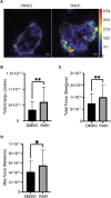
R491-treatment in BJ-Ras cells induces cellular contractile forces. (A) Representative traction force maps of control- and R491-treated cells with the color showing the magnitude of traction force (in pascals), as indicated in the color key. Quantification of (B) the total traction forces (traction forces integrated over cell area) exerted by the cells, (C) the total strain energy and the (D) maximum force is shown, as indicated. Traction force microscope measurements were taken from 5 µM DMSO- or R491-treated cells, from three biological repeats, n = 55 and n = 42 cells respectively. Data presented as mean ± SD. *p ≤ 0.05, **p ≤ 0.01. Scale bars: 10 µm.
FIGURE 8
FIGURE 8
R491 increases the fraction and number of small focal adhesions. (A) Representative confocal microscopy images of BJ-Ras cells treated with 5 μM control or R491, showing vimentin, phospho-tyrosine, F-actin, as indicated, and merged images with vimentin (red), phospho-tyrosine (yellow), actin (green) and nuclei (blue). Inset images: magnified views of boxed areas in the larger images. Scale bars: 10 μm. The graphs show (B) the average size of cell-matrix adhesions/per cell, with n = 516 and 478 focal adhesions for control- and R491-treated cells, respectively, and (C) the frequency distribution of the binned sizes of focal adhesions of control- (black) or R491- (grey) treated cells. **p ≤ 0.01. The difference in the frequency distribution was significant with p ≤ 0.0001.

References

    1. Abercrombie M. (1961). The bases of the locomotory behaviour of fibroblasts. Exp. Cell Res. 8, 188–198. 10.1016/0014-4827(61)90348-2 - DOI - PubMed
    1. Beningo K. A., Dembo M., Kaverina I., Small J. V., Wang Y. L. (2001). Nascent focal adhesions are responsible for the generation of strong propulsive forces in migrating fibroblasts. J. Cell Biol. 153, 881–888. 10.1083/jcb.153.4.881 - DOI - PMC - PubMed
    1. Bollong M. J., Pietila M., Pearson A. D., Sarkar T. R., Ahmad I., Soundararajan R., et al. (2017). A vimentin binding small molecule leads to mitotic disruption in mesenchymal cancers. Proc. Natl. Acad. Sci. U. S. A. 114, E9903–E9912. 10.1073/pnas.1716009114 - DOI - PMC - PubMed
    1. Cheng F., Shen Y., Mohanasundaram P., Lindstrom M., Ivaska J., Ny T., et al. (2016). Vimentin coordinates fibroblast proliferation and keratinocyte differentiation in wound healing via TGF-beta-Slug signaling. Proc. Natl. Acad. Sci. U. S. A. 113, E4320–E4327. 10.1073/pnas.1519197113 - DOI - PMC - PubMed
    1. Chernyatina A. A., Nicolet S., Aebi U., Herrmann H., Strelkov S. V. (2012). Atomic structure of the vimentin central alpha-helical domain and its implications for intermediate filament assembly. Proc. Natl. Acad. Sci. U. S. A. 109, 13620–13625. 10.1073/pnas.1206836109 - DOI - PMC - PubMed