. 2022 Dec 9;50(22):12896-12912.
doi: 10.1093/nar/gkac1146.

The Caulobacter crescentus DciA promotes chromosome replication through topological loading of the DnaB replicative helicase at replication forks

Affiliations

The Caulobacter crescentus DciA promotes chromosome replication through topological loading of the DnaB replicative helicase at replication forks

Shogo Ozaki et al. Nucleic Acids Res. .

Abstract

The replicative DNA helicase translocates on single-stranded DNA to drive replication forks during chromosome replication. In most bacteria the ubiquitous replicative helicase, DnaB, co-evolved with the accessory subunit DciA, but how they function remains incompletely understood. Here, using the model bacterium Caulobacter crescentus, we demonstrate that DciA plays a prominent role in DNA replication fork maintenance. Cell cycle analyses using a synchronized Caulobacter cell population showed that cells devoid of DciA exhibit a severe delay in fork progression. Biochemical characterization revealed that the DnaB helicase in its default state forms a hexamer that inhibits self-loading onto single-stranded DNA. We found that upon binding to DciA, the DnaB hexamer undergoes conformational changes required for encircling single-stranded DNA, thereby establishing the replication fork. Further investigation of the functional structure of DciA revealed that the C-terminus of DciA includes conserved leucine residues responsible for DnaB binding and is essential for DciA in vivo functions. We propose that DciA stimulates loading of DnaB onto single strands through topological isomerization of the DnaB structure, thereby ensuring fork progression. Given that the DnaB-DciA modules are widespread among eubacterial species, our findings suggest that a common mechanism underlies chromosome replication.


Figures

Figure 1.
Figure 1.
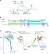
A model for replicative DNA helicase loading. (A) A model for replicative helicase loading in E. coli. The replicative DnaBEC helicase (blue circles) forms a hexameric, closed ring as a default state. At the replication origin or stalled replication forks, the DnaBEC hexamer undergoes conformational changes to open the ring structure, thereby allowing it to engage ssDNA. (B) Comparison of DnaBEC and C. crescentus DnaB. The two sequences were aligned using ClustalW and are shown schematically. The overall similarity at the amino acid level was 52%. (C) Structural comparison among E. coli DnaC (PDBID 6QEL, Chain G), phage lambda P (PDBID 6BBM, Chain W), and a predicted C. crescentus DciA structure generated by AlphaFold. The central DANL domain (amino acids 47–123; DUF721 in Pfam), the N-terminal extension (amino acids 1–46), and the C-terminal extension (amino acids 124–179) are highlighted in different colors. Previously determined E. coli DnaB binding regions are indicated by dotted circles. The different domains of DciA are highlighted in different colors.
Figure 2.
Figure 2.
DciA is essential for C. crescents proliferation (A) Schematic representation of a DciA-depletable strain (SHQ209), in which the xylose-dependent promoter (PxylX) flanking the E. coli rrnB T1T2 transcriptional terminator (rrnB_ter) is integrated at the dciA promoter locus. The relative positions of Cori, dnaA, dciA, and dnaB are indicated on the circular C. crescentus genome. (B) Colony formation in the PxylX-dciA strain. The wild-type NA1000 (WT) and SH209 strains were grown overnight at 30°C in PYE medium supplemented with 0.1% xylose. Five-fold serial dilutions of the overnight culture were spotted on PYE agar supplemented with 0.1% xylose or 0.1% glucose, followed by incubation at 30°C for 2d. When the SHQ209 strain harboring pMR10 (vector) or pMR10-HAdciA (dciA) was analyzed, kanamycin was added to the medium. (C) Cell morphology and DNA content of the PxylX-dciA strain. The SHQ209 strain harboring pMR10 (vector) or pMR10-HAdciA (dciA) was grown for 9 h at 30°C in PYE medium supplemented with 0.1% glucose and kanamycin, followed by fixation in 70% ethanol. After DNA staining with SYTOX Green, cell morphology and DNA content were analyzed using phase-contrast microscopy and flow cytometry, respectively. (D) The distributions of cell lengths are shown using a box plot. The p-value was calculated using the Mann–Whitney–Wilcoxon test.
Figure 3.
Figure 3.
DciA is important for fork progression (A) Schematic representation of a DciA-degradable strain (SHQ258), in which the native dciA gene is replaced with a dciA-DAS + 4 fusion, and the E. coli sspB gene (SspBec) is integrated downstream of the xylose dependent promoter (PxylX) at the xylX locus. Upon induction of SspBec with 0.1% xylose, DAS + 4-tagged DciA is degraded by the ClpXP protease. The relative positions of Cori, dnaA, dciA and xylX are indicated on the circular C. crescentus genome. (B) DNA replication activity in the absence of DicA. SHQ254 (NA1000 xylX::sspBEC) and its derivatives, DnaA-degradable SHQ259 (NA1000 xylX::sspBEC dnaA::DAS + 4) and DciA-degradable SH258 (NA1000 xylX::sspBEC dciA::DAS + 4), cells were grown exponentially in PYE medium supplemented with 0.1% glucose. After addition of 0.1% xylose, cells were further incubated for 20 min at 30°C, followed by cell cycle synchronization. The harvested G1 phase cells were incubated at 30°C in PYE medium supplemented with 0.1% xylose. A portion of the cells was withdrawn at the indicated time, fixed in 70% ethanol, and analyzed by flow cytometry. The DNA profiles of the control SHQ254 cells are colored in red and overlaid with those of SHQ259 and SHQ258. (C) DNA replication activity upon overexpression of DciA. Wild-type NA1000 cells harboring pQF (vector) or pQFdciA (DciA) were grown exponentially at 30°C in PYE medium supplemented with tetracycline. After addition of the inducer, cumate (1 μM), cells were further incubated for 20 min at 30°C, followed by cell cycle synchonization. The harvested G1 phase cells were incubated at 30°C for 0–150 min in PYE medium supplemented with 10 μM cumate, 20 μg/ml cephalexin, and tetracycline. A portion of the cells were withdrawn at the indicated time, fixed in 70% ethanol, and analyzed by flow cytometry. The DNA profiles of cells with the vector control are colored in red and overlaid with those of cells with pQFdciA.
Figure 4.
Figure 4.
DciA stimulates ATP hydrolysis by DnaB. (A) Size exclusion chromatography of DnaB and DciA. DnaB-His (10 nmol; top), His-DciA (25 nmol; middle), or a mixture of both (10 nmol of DnaB-His and 25 nmol of His-DciA; bottom) were separated using a Superdex 200 column. The elution fractions were analyzed by SDS–15% PAGE and Coomassie brilliant blue staining. The elution positions of the molecular weight marker proteins are indicated. To deduce the stoichiometry of DnaB-His per His-DciA in the elution fractions (lanes 2 and 3) for the DnaB-His/His-DciA mixture, the same samples were reanalyzed by SDS–15% PAGE and Coomassie brilliant blue staining (bottom). The indicated amounts of DnaB-His and His-DciA proteins were used as quantitative standard. (BG) ATPase assay. [α-32P]ATP was incubated with DnaB-His at 30°C for 10 min, followed by thin layer chromatography (TLC). A representative chromatograph (B) and kinetic analysis (C) for a titration of DnaB-His (2.1, 4.2, 8.3, 17, or 33 nM as hexamer) in the presence of 0.1 mM [α-32P]ATP were shown. WT, wild type. K234A, an ATPase-dead variant. For panel D and E, ATP hydrolysis of DnaB-His (8.3 nM as hexamer; 50 nM as monomer) was analyzed using TLC at various [α-32P]ATP concentrations (5–160 μM) in the presence or absence of His-DciA (100 nM as monomer) (D) and the ATPase rate was fit using Michaelis-Menten kinetics in JMP statistical software (https://www.jmp.com). For panel F and G, ATP hydrolysis was analyzed in buffer containing [α-32P]ATP (100 μM), DnaB-His (0 or 8.3 nM as hexamer), HisDciA (0 or 100 nM as monomer), and 76-mer ssDNA (0, 3.1, 6.3, 13 or 25 nM of oligo 764).
Figure 5.
Figure 5.
DciA stimulates the DnaB helicase activity DNA helicase assay using a forked DNA substrate. A schematic of the assay is shown (A). The bottom strand of the forked DNA substrate is 5′-FAM labelled (green circles) and hybridizes with competitor ssDNA upon separation from the upper strand of the forked DNA substrate. The substrate DNA (12.5 nM) was incubated at 30°C for 0–30 min in buffer containing competitor ssDNA (62.5 nM) and the indicated concentrations of DnaB-His and His-DciA. The products were separated using 6% polyacrylamide gel electrophoresis. Reaction curves and representative gel images for a DciA-titration experiment (B) and a time-course experiment (C) are shown.
Figure 6.
Figure 6.
DciA stimulates loading of the DnaB helicase onto the bubble DNA structure (AB) A bubble DNA substrate (12.5 nM) was incubated at 30°C for 20 min in buffer containing 5′-FAM labelled competitor ssDNA (62.5 nM) and the indicated concentrations of the recombinant proteins. The products were separated using 9% polyacrylamide gel electrophoresis (A). For panel B, 32P-labelled DNA substrate and cold competitor were used instead of cold DNA substrate and 5′-FAM labelled competitor. In both cases, separation of the bubble DNA substrate allows the bottom strand to hybridize with competitor ssDNA. A forked DNA substrate was used as control. A schematic of the assay is shown above the gel image. The 5′-DNA ends labelled by FAM and 32P were indicated by green circles and red circles, respectively. (CD) His-DciA titration experiments in the presence of DnaB-His (16 nM as hexamer). A representative gel image is shown (C). Scatter plots generated by multiple independent reactions (n > 3) are used to draw reaction curves (D). The number of points reflects the exact sample size. Intermediates of the rection were indicated (see Supplementary Figure S5 for details). (E) His-DciA titration experiments with DNA substrates bearing a different bubble size (18, 28 or 38-mer). A representative gel image was shown in Supplementary Figure S6C. (F) DnaC titration experiments in the presence or absence of E. coli DnaBEC (100 nM). (GH) The interplay between His-DciA and E. coli DnaBEC. The activities of DnaB-His and E. coli DnaBEC were assessed in the presence of HisDciA or DnaC (100 nM). A representative gel (F) and bar graphs (G) with scatter plots generated by multiple independent reactions (n > 3) are shown. The number of points reflects the exact sample size.
Figure 7.
Figure 7.
The DciA C-terminus operates DnaB (A) Schematic representation of DciA truncation constructs. sfTq2 proteins fused to the N-terminus of full-length DciA (sfTq2-DciA), or the truncated versions (N, 1–47 aa; DANL, 46–127 aa; C, 124–179 aa) were used for the pulldown assays. For plasmid complementation assays, pMR10-dciA derivatives carrying the dciA allele (Δ9, 1–170 aa; Δ19, 1–160 aa) were used. The schematic shows the predicted secondary structures (alpha helices in blue and beta strands in orange). Multiple sequence alignments were created by Protein BLAST search for DciA and the top 500 sequence data were used to generate Weblogo representation (66). (B) E. coli crude extracts with and without sfTq2-DciA were incubated in the presence (+) or absence (–) of DnaB-His, followed by pulldown using Ni-conjugated magnetic beads. After washing, materials retained on the beads were analyzed using SDS–12% PAGE and Coomassie Brilliant Blue staining. AS, ammonium sulfate-precipitated sfTq2-DciA; Input, a mixture of AS and DnaB-His; Pulldown, an elution fraction. (C) Plasmid complementation test. pMR10-HAdciA derivatives (Δ9 and Δ19) or pMR10dciA derivatives with the indicated dciA allele were analyzed as described in Figure 2D. (D) Helicase assay. Wild-type His-DciA or His-DciA(L167S) were analyzed as described in Figure 5. Reaction curves and representative gel images for a DciA-titration experiment are shown.

Similar articles

Cited by

References

    1. Fernandez A.J., Berger J.M.. Mechanisms of hexameric helicases. Crit. Rev. Biochem. Mol. Biol. 2021; 56:621–639. - PMC - PubMed
    1. O’Donnell M.E., Li H.. The ring-shaped hexameric helicases that function at DNA replication forks. Nat. Struct. Mol. Biol. 2018; 25:122–130. - PMC - PubMed
    1. Bell S.P., Kaguni J.M.. Helicase loading at chromosomal origins of replication. Cold Spring Harb. Perspect. Biol. 2013; 5:a010124. - PMC - PubMed
    1. Katayama T., Ozaki S., Keyamura K., Fujimitsu K.. Regulation of the replication cycle: conserved and diverse regulatory systems for DnaA and oriC. Nat. Rev. Microbiol. 2010; 8:163–170. - PubMed
    1. Kaguni J.M. Replication initiation at the Escherichiacoli chromosomal origin. Curr. Opin. Chem. Biol. 2011; 15:606–613. - PMC - PubMed

Publication types

MeSH terms