. 2023 Mar 21;147(12):956-972.
doi: 10.1161/CIRCULATIONAHA.122.061934. Epub 2022 Dec 9.

Placental Inflammation Leads to Abnormal Embryonic Heart Development

Affiliations

Placental Inflammation Leads to Abnormal Embryonic Heart Development

Eleanor J Ward et al. Circulation. .

Abstract

Background: Placental heart development and embryonic heart development occur in parallel, and these organs have been proposed to exert reciprocal regulation during gestation. Poor placentation has been associated with congenital heart disease, an important cause of infant mortality. However, the mechanisms by which altered placental development can lead to congenital heart disease remain unresolved.

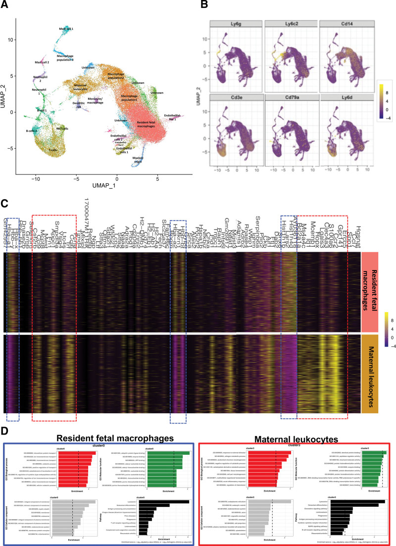
Methods: In this study, we use an in vivo neutrophil-driven placental inflammation model through antibody depletion of maternal circulating neutrophils at key stages during time-mated murine pregnancy: embryonic days 4.5 and 7.5. Pregnant mice were culled at embryonic day 14.5 to assess placental and embryonic heart development. A combination of flow cytometry, histology, and bulk RNA sequencing was used to assess placental immune cell composition and tissue architecture. We also used flow cytometry and single-cell sequencing to assess embryonic cardiac immune cells at embryonic day 14.5 and histology and gene analyses to investigate embryonic heart structure and development. In some cases, offspring were culled at postnatal days 5 and 28 to assess any postnatal cardiac changes in immune cells, structure, and cardiac function, as measured by echocardiography.

Results: In the present study, we show that neutrophil-driven placental inflammation leads to inadequate placental development and loss of barrier function. Consequently, placental inflammatory monocytes of maternal origin become capable of migration to the embryonic heart and alter the normal composition of resident cardiac macrophages and cardiac tissue structure. This cardiac impairment continues into postnatal life, hindering normal tissue architecture and function. Last, we show that tempering placental inflammation can prevent this fetal cardiac defect and is sufficient to promote normal cardiac function in postnatal life.

Conclusions: Taken together, these observations provide a mechanistic paradigm whereby neutrophil-driven inflammation in pregnancy can preclude normal embryonic heart development as a direct consequence of poor placental development, which has major implications on cardiac function into adult life.

Keywords: fetus; heart defects, congenital; inflammation; macrophages; mothers; neutrophils; placenta.


Conflict of interest statement

None.

Figures

Figure 1.
Figure 1.
An NDPI model. Neutrophils were depleted at days 4.5 and 7.5 of pregnancy with αLy6G. Mice were euthanized at embryonic day E14.5 of pregnancy, and placentas were harvested to assess the structure and immune composition. Isotype control treated (referred to hereafter as control; white) and neutrophil depleted (referred to hereafter as neutrophil-driven placental inflammation [NDPI]; pink). A, Expression of activation markers by maternal blood neutrophils. B, CCR2 expression on maternal blood monocytes. C, ELISA showing the concentration of interleukin-1β (IL-1β) and tumor necrosis factor-α (TNF-α) in plasma and placenta digest supernatants. D, Immunofluorescent staining of placentas with CD45 (red) and wheat germ agglutinin (WGA; green) and cell nuclei with DAPI (blue). Expression is shown in the decidual layer (left) and labyrinth layer (right). E through G, Neutrophil subpopulations from placentas analyzed by flow cytometry expressed as absolute cell number per gram of tissue. E, CD11b+Ly6G+ neutrophils, (F) CD11b+Ly6G+ TNF-α+ neutrophils, and (G) neutrophils expressing CD11b+Ly6G+ CXCR2, CD11b+Ly6G+ CD114+, and CD11b+Ly6G+ MMP9+. H, Macrophages from placentas analyzed by flow cytometry expressed as absolute cell number per gram of tissue. I, Inflammatory monocytes from placentas analyzed by flow cytometry expressed as absolute cell number per gram of tissue. J, F4/80Ly6Chi populations from placentas were analyzed for the expression of major histocompatibility complex II (MHCII), CCR2, CXCR2, CCR5, and CCR6, expressed as median fluorescent intensity. Representative histograms are shown below the quantification (gray=control, pink=NDPI). Each symbol represents an individual mouse from different pregnancies, and statistical significance was tested by unpaired Student t test or Welch t test (G [CXCR2 and CD114] and H). In all cases, data are mean±SEM. MFI indicates median fluorescence intensity; MMP9, matrix metalloproteinase-9; and ns, not significant. *P≤0.05. **P≤0.01. ***P≤0.001. ****P≤0.0001.
Figure 2.
Figure 2.
Neutrophil-driven inflammation promotes a breakdown in the placental tissue barrier. A through E, Neutrophils were depleted at days 4.5 and 7.5 of pregnancy with αLy6G. Mice were euthanized at E14.5 of pregnancy, and placentas were harvested. Control (white) and neutrophil-driven placental inflammation (NDPI; pink). A through C, RNA sequencing from placentas. A, Volcano plot showing differentially expressed genes in control vs NDPI placentas. B, Top 15 modulated pathways in placentas of NDPI mice compared with control. C, Heat map showing gene expression of extracellular matrix components of NDPI or control placentas. D, Reverse transcriptase–polymerase chain reaction showing the gene expression of Col1a1, Col1a2, Col4a6, Col5a1, Col9a1, Col9a2, and Col9a3. E, Immunofluorescent images of control or NDPI murine placentas stained with DAPI (blue), collagen (Col) IV (red), and isolectin B4 (red). Magnified images of the decidua and labyrinth of the placenta and graphs showing the quantification of Col IV staining. F and G, Immunofluorescent staining of term placentas from women with healthy, preeclamptic pregnancies or pregnancies carrying babies with congenital heart disease (CHD) for (F) smooth muscle actin (SMA; green), (G) Col I (green; top), and Col IV (orange; bottom). In all images, wheat germ agglutinin (WGA) staining is in red, and cell nuclei are stained with DAPI (blue). Quantification of area stained with SMA, Col I, and Col IV is shown below images. Each symbol represents an individual sample from different pregnancies, and statistical significance was tested by unpaired Student t test or Welch t test (E) or 1-way ANOVA (F and G). In all cases, data are mean±SEM. ns Indicates not significant. *P≤0.05. **P≤0.01. **P≤0.001.
Figure 3.
Figure 3.
Placental inflammation leads to poor embryonic cardiac development. Neutrophils are depleted at days 4.5 and 7.5 of pregnancy with αLy6G. Mice were euthanized at E14.5 of pregnancy, and embryos were harvested. Control is in white; neutrophil-driven placental inflammation (NDPI) is in pink. A, Immunofluorescent images of cross section of embryonic day (E) 14.5 embryo heart. Sections were stained with DAPI (blue), endomucin (EMCN; green), isolectin B4 (ISOb4; yellow), and actinin-2 (ACTN2; red). B, High-resolution episcopic microscopy images of E14.5 hearts. C,Quantification of the in vivo uptake of BrdU into CD45CD31+ endothelial cells and CD45troponin-T+ cardiomyocytes in embryo hearts. Each symbol represents an individual mouse, and statistical significance was tested by unpaired Student t test. In all cases, data are mean±SEM. ns Indicates not significant. *P≤0.05. **P≤0.01. ***P≤0.001. ****P≤0.0001.
Figure 4.
Figure 4.
CCR2-driven accumulation of maternal proinflammatory leukocytes in embryonic hearts of NDPI pregnancies. Neutrophils were depleted at days 4.5 and 7.5 of pregnancy with αLy6G. Mice were euthanized at embryonic day (E)14.5 of pregnancy, and hearts were dissected from harvested embryos. Control is shown in white, and neutrophil depleted (neutrophil-driven placental inflammation [NDPI]) is shown in pink. A, Flow cytometry plots of leukocytes in E14.5 fetal hearts and NDPI pregnancies; CD45.1+ CD45.2 cells are of maternal origin and CD45.1+CD45.2+ cells are of fetal origin. Graphs show quantification of number of maternal cells per embryo heart or embryo liver. B, Flow cytometry quantification of different maternal leukocyte subsets in embryo hearts. C, Median fluorescent intensity of major histocompatibility complex II (MHCII) and CCR2 on maternal F4/80+Ly6Chi cells found in embryo hearts. Bottom, Representative histograms. D, Immunofluorescent images of cross section of E14.5 embryo heart from CCR2+/−control and CCR2+/−NDPI pregnancies. Sections were stained with DAPI (blue), endomucin (EMCN; green), isolectin B4 (ISOb4; yellow), and actinin-2 (ACTN2; red). Graph indicates quantification of endomucin staining of embryonic hearts expressed as percent of total heart area. E, Quantification of the in vivo uptake of BrdU into CD45CD31+ endothelial cells and CD45troponin-T+ cardiomyocytes in embryo hearts from CCR2+/−control and CCR2+/−NDPI pregnancies compared with control. F, Quantification of absolute numbers of CX3CR1 fetal cells in E14.5 embryonic hearts from control, NDPI, CCR2+/−control, and CCR2+/−NDPI pregnancies. G, Proportion of CX3CR1+CCR2+ and CX3CR1+CCR2 fetal cells in E14.5 embryonic hearts from control, NDPI, CCR2+/−control, and CCR2+/−NDPI pregnancies. H, Absolute number of CX3CR1+CCR2+ fetal cells in E14.5 embryonic hearts from control, NDPI, CCR2+/−control, and CCR2+/−NDPI pregnancies. Each symbol represents an individual mouse, and statistical significance was tested by unpaired Student t test. In all cases, data are mean±SEM. MFI indicates median fluorescence intensity; and ns, not significant. *P≤0.05. **P≤0.01. ***P≤0.001. ****P≤0.0001 (A–C): 1-way ANOVA with Dunnett multiple-comparison test compared with control (D–F and H); Brown-Forsythe ANOVA test with Dunnett multiple-comparison test (E, troponin-T); or 2-ANOVA comparing means of CCR2+ or CCR2 percent between mouse groups with the Tukey multiple-comparison test (G).
Figure 5.
Figure 5.
Single-cell sequencing of leukocyte s from E14.5 embryo hearts. Neutrophils were depleted at days 4.5 and 7.5 of pregnancy with αLy6G. Mice were sacrificed at embryonic day (E) 14.5 of pregnancy, and hearts were dissected from harvested embryos from control or NDPI pregnancies. CD45+ cells were isolated from heart single cell suspensions with CD45 preeclamptic and antipreeclamptic microbeads. Single-cell sequencing was performed on the isolated cells. A, Uniform Manifold Approximation and Projection showing cell clusters found in hearts of fetuses from control and NDPI pregnancies. B, Feature plots showing expression of key genes within clusters. C, Heart map analyses comparing gene expression between resident fetal macrophage and maternal leukocyte clusters. D, Enrichment analysis of Gene Ontology (GO) terms and pathways for differentially expressed genes. GO analysis including biological process, cellular component, and molecular function. Pathway analysis based on the KEGG database.
Figure 6.
Figure 6.
Aberrant embryonic cardiac development from NDPI pregnancies continues into postnatal life. Neutrophils were depleted at days 4.5 and 7.5 of pregnancy with αLy6G. Offspring of these dams were euthanized at postnatal day (P) 5 or P28 as indicated, and heart structure and immune composition were assessed. Offspring from control (white) and neutrophil-driven placental inflammation (NDPI; pink). A, P28 offspring body weights in grams and heart:body weight ratio from control, NDPI, CCR2+/−control, and CCR2+/−NDPI pregnancies. B, Representative echocardiography plots of P28 hearts of offspring from control, NDPI, CCR2+/−control, and CCR2+/−NDPI pregnancies. C,Graphs showing quantification of heart parameters determined by echocardiography. D, Flow cytometric quantification of total CD45+ leukocytes from P28 offspring hearts of control, NDPI, CCR2+/−control, and CCR2+/−NDPI pregnancies. E, Flow cytometric analyses of macrophage and monocyte populations as determined by F4/80 and Ly6C staining in P28 offspring hearts from control, NDPI, CCR2+/−control, and CCR2+/−NDPI pregnancies. F, Proportion of macrophage and monocyte populations shown in E in P28 offspring hearts from control, NDPI, CCR2+/−control, and CCR2+/−NDPI pregnancies. G, Absolute number per gram of heart tissue of monocyte and macrophage populations shown in E in P28 offspring hearts from control, NDPI, CCR2+/−control, and CCR2+/−NDPI pregnancies. H, Percentage of F4/80+Ly6C+ MHCII+ (left) and F4/80+Ly6C+ CCR2+ in P28 offspring hearts from control, NDPI, CCR2+/−control, and CCR2+/−NDPI pregnancies. Each symbol represents an individual mouse, and statistical significance was tested by 1-way ANOVA with the Dunnett multiple-comparison test or Brown-Forsythe ANOVA test with Dunnett post hoc comparison (G, F4/80+Ly6C+) compared with control. In all cases, data are mean±SEM. LV indicates left ventricular; and ns, not significant. *P≤0.05. **P≤0.01. ***P≤0.001. ****P≤0.0001.
Figure 7.
Figure 7.
Quelling placental inflammation prevents abnormal cardiac development. Neutrophils were depleted at days 4.5 and 7.5 of pregnancy with αLy6G. At day 8.5 of pregnancy, tumor necrosis factor-α (TNF-α) was neutralized by injecting aTNF-α immunoglobulin G (IgG) intravenously. Mice were euthanized at embryonic day (E) 14.5 of pregnancy, and placentas and embryos were harvested to assess the structure and immune composition. Control is shown in white, neutrophil-driven placental inflammation (NDPI) in pink, and neutrophil-depleted and TNF-α neutralized (aTNF-αNDPI) in blue. A, Immunofluorescent images of cross section of E14.5 embryo heart. Sections were stained with DAPI (blue), wheat germ agglutinin (WGA; red), and smooth muscle actin (SMA; green). Graph showing ventricular wall thickness quantification. B, Hematoxylin and eosin sections of E14.5 embryonic hearts from aTNF-αNDPI pregnancies compared with control Graph represents left ventricular wall thickness. C, Immunofluorescent images showing the expression of endomucin (orange) and WGA (green) in cross sections of hearts from postnatal day (P) 5. Scale bar, 1 mm. Magnified images showing endomucin staining of the ventricle. Graph showing the proportion of total CD31+ cells in P5. D, Echocardiography plot with graphs (bottom) showing quantification of heart parameters determined by echocardiography. Each symbol represents an individual mouse, and statistical significance was tested by 1-way ANOVA with Bonferroni post hoc correction. In all cases, data are mean±SEM. ns Indicates not significant. *P≤0.05. **P≤0.01. ***P≤0.001. ****P≤0.0001.

Comment in

References

    1. Jorgensen M, McPherson E, Zaleski C, Shivaram P, Cold C. Stillbirth: the heart of the matter. Am J Med Genet A. 2014;164:691–699. doi: 10.1002/ajmg.a.36366 - PubMed
    1. National Congenital Anomaly and Rare Disease Registration Service . NCARDRS congenital anomaly statistics 2017. PHE publications gateway number GW-473. 2017. Accessed December 13, 2022. https://www.gov.uk/government/publications/ncardrs-congenital-anomaly-an...
    1. Buijtendijk MFJ, Barnett P, van den Hoff MJB. Development of the human heart. Am J Med Genet C Semin Med Genet. 2020;184:7–22. doi: 10.1002/ajmg.c.31778 - PMC - PubMed
    1. Doubilet PM, Benson CB. Embryonic heart rate in the early first trimester: what rate is normal? J Ultrasound Med. 1995;14:431–434. doi: 10.7863/jum.1995.14.6.431 - PubMed
    1. Burton GJ, Jauniaux E. Development of the human placenta and fetal heart: synergic or independent? Front Physiol. 2018;9:373. doi: 10.3389/fphys.2018.00373 - PMC - PubMed

Publication types