. 2023 Feb;27(2):21.
doi: 10.3892/mmr.2022.12908. Epub 2022 Dec 9.

G9a inhibition promotes the formation of pacemaker-like cells by reducing the enrichment of H3K9me2 in the HCN4 promoter region

Affiliations

G9a inhibition promotes the formation of pacemaker-like cells by reducing the enrichment of H3K9me2 in the HCN4 promoter region

Pei Xu et al. Mol Med Rep. 2023 Feb.

Abstract

Biological pacemakers, made of pacemaker-like cells, are promising in the treatment of bradyarrhythmia; however, the inefficiency of stem cell differentiation into pacemaker-like cells has limited their clinical application. Previous studies have reported that histone H3 at lysine 9 (H3K9) methylation is widely involved in the proliferation and differentiation of cardiomyocytes, but the specific role of H3K9 dimethylation (H3K9me2) in the formation of pacemaker cells remains unclear. The present study evaluated the functional role of H3K9me2 in the differentiation of bone marrow mesenchymal stem cells (BMSCs) into pacemaker-like cells. Rat BMSCs pretreated with the euchromatic histone lysine methyltransferase 2 (G9a) inhibitor BIX01294 were transfected with a T-box 18 overexpression plasmid to induce BMSCs to form pacemaker-like cells. The induced pacemaker-like cells were analyzed using reverse transcription-quantitative PCR (RT-qPCR) and immunofluorescence to assess the efficiency of differentiation. The enrichment of H3K9me2 in the hyperpolarized-activated cyclic nucleotide-gated cation channel (HCN)4 promoter region was assessed by chromatin immunoprecipitation (ChIP). In addition, BIX01294 was injected into rats, and the protein and mRNA expression levels of HCN4 were assessed using western blotting and RT-qPCR. After interference with G9a using BIX01294, ChIP results demonstrated that H3K9me2 levels in the promoter region of HCN4 were markedly decreased. Immunofluorescence and RT-qPCR demonstrated that the protein expression levels of certain cardio-specific proteins in the treated group were significantly higher compared with those in the untreated group. In vivo experiments demonstrated that interference with G9a could cause pathological hypertrophy. Furthermore, in vitro and in vivo inhibition of G9a could increase the differentiation and proliferation of pacemaker-like cells by decreasing the levels of H3K9me2 in the promoter region of HCN4 gene.

Keywords: euchromatic histone lysine methyltransferase 2; histone H3 at lysine 9; hyperpolarized-activated cyclic nucleotide-gated cation channel 4; pacemaker-like cells; promoter region.


Conflict of interest statement

The authors declare that they have no competing interests.

Figures

Figure 1.
Figure 1.
Characteristics of BMSCs and transfection efficiency. (A) Fluorescence-activated cell sorting analysis of CD29, CD44, CD34 and CD45 protein expression levels in BMSCs. (B) Cellular morphological features of BMSCs, which exhibited a spindle shape. Scale bar, 50 µm. (C) Red fluorescence of BMSCs after transfection with OE-Tbx18 for 48 h. Scale bar, 50 µm. (D) The microscopic morphology of BMSCs gradually evolved into strip-like features after 10 days of transduction by OE-Tbx18. Scale bar, 50 µm. (E) Western blotting of Tbx18 protein expression levels after transfection for 72 h. BMSCs without transfection were used as the control, BMSCs transfected with the empty vector were used as the NC. (F) Reverse transcription-quantitative PCR analysis of Tbx18 mRNA expression levels after transfection for 72 h. **P<0.01. BMSCs, bone marrow mesenchymal stem cells; OE-Tbx18, T-box 18 overexpression plasmid; NC, negative control.
Figure 2.
Figure 2.
Immunostaining, western blotting and reverse transcription-quantitative PCR analysis of target protein and mRNA expression levels after transfection. (A) Protein expression levels of cTnI in BMSCs were assessed using immunostaining with cTnI-specific primary antibodies and a Cy3 secondary antibody. Scale bar, 50 µm. (B) Quantitative analysis of the relative mRNA expression levels of HCN4, α-SA and cTnI. (C) Western blotting demonstrated increased HCN4, α-SA and cTnI protein expression levels. BMSCs without transfection were used as the control and BMSCs transfected with the empty vector were used as the NC. **P<0.01. cTnI, cardiac troponin I; HCN4, hyperpolarization-activated cyclic nucleotide-gated channel 4; α-SA, α-striated actin; NC, negative control; CM, cardiomyocyte; BMSCs, bone marrow mesenchymal stem cells.
Figure 3.
Figure 3.
Expression of target protein after G9a inhibition treatment. (A) Western blotting demonstrated increased HCN4, α-SA and cTnI protein expression levels in different groups. (B) Quantitative analysis of the mRNA expression levels of HCN4, α-SA and cTnI. (C) Chromatin immunoprecipitation-qPCR analysis for H3K9me2 binding to the HCN4 promoter. The histogram showed the amount of immunoprecipitated DNA as detected using the qPCR assay. Values are indicated as % of input. *P<0.05 and **P<0.01. cTnI, cardiac troponin I; HCN4, hyperpolarization-activated cyclic nucleotide-gated channel 4; α-SA, α-striated actin; CM, cardiomyocyte; BIX, H3K9me2, histone H3 at lysine 9 dimethylation; Tbx18, T-box 18 overexpression plasmid.
Figure 4.
Figure 4.
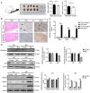
BIX01294 promotes cardiac hypertrophy. (A) BIX01294 was injected through the caudal vein, and heart volume and weight were assessed at day 50. (B) H&E staining of the heart and immunohistochemistry of sinoatrial node tissue. The arrow indicated positive cells. Scale bar, 50 µm. (C) Quantitative analysis of the mRNA expression levels of Nppa, Nppb and Myh7. (D) Western blotting was used to assess the protein expression levels of H3K9me2, G9a, Nppa, Nppb and Myh7. *P<0.05 and **P<0.01. H3K9me2, histone H3 at lysine 9 dimethylation; G9a, euchromatic histone lysine methyltransferase 2; H&E, haematoxylin and eosin; Nppa, natriuretic peptide A; Nppb, natriuretic peptide B; Myh7, myosin heavy chain 7.

Similar articles

Cited by

References

    1. Sun X, Li H, Zhu Y, Xu P, Zuo Q, Li B, Gu X. 5-Azacytidine-induced cardiomyocyte differentiation of very small embryonic-like stem cells. Stem Cells Int. 2020;2020:5162350. doi: 10.1155/2020/5162350. - DOI - PMC - PubMed
    1. Farraha M, Kumar S, Chong J, Cho HC, Kizana E. Gene therapy approaches to biological pacemakers. J Cardiovasc Dev Dis. 2018;5:50. doi: 10.3390/jcdd5040050. - DOI - PMC - PubMed
    1. Li Y, Yang M, Zhang G, Li L, Ye B, Huang C, Tang Y. Transcription factor TBX18 promotes adult rat bone mesenchymal stem cell differentiation to biological pacemaker cells. Int J Mol Med. 2018;41:845–851. - PMC - PubMed
    1. Xiao H, Yang YJ, Lin YZ, Peng S, Lin S, Song ZY. Transcription factor Tbx18 induces the differentiation of c-kit+ canine mesenchymal stem cells (cMSCs) into SAN-like pacemaker cells in a co-culture model in vitro. Am J Transl Res. 2018;10:2511–2528. - PMC - PubMed
    1. Gorabi AM, Hajighasemi S, Tafti HA, Atashi A, Soleimani M, Aghdami N, Saeid AK, Khori V, Panahi Y, Sahebkar A. TBX18 transcription factor overexpression in human-induced pluripotent stem cells increases their differentiation into pacemaker-like cells. J Cell Physiol. 2019;234:1534–1546. doi: 10.1002/jcp.27018. - DOI - PubMed

MeSH terms

Substances