. 2023 Feb 6;155(2):e202213173.
doi: 10.1085/jgp.202213173. Epub 2022 Dec 9.

Acid-sensing ion channel 1a activates IKCa/SKCa channels and contributes to endothelium-dependent dilation

Affiliations

Acid-sensing ion channel 1a activates IKCa/SKCa channels and contributes to endothelium-dependent dilation

Selina M Garcia et al. J Gen Physiol. .

Abstract

Acid-sensing ion channel 1a (ASIC1a) belongs to a novel family of proton-gated cation channels that are permeable to both Na+ and Ca2+. ASIC1a is expressed in vascular smooth muscle and endothelial cells in a variety of vascular beds, yet little is known regarding the potential impact of ASIC1a to regulate local vascular reactivity. Our previous studies in rat mesenteric arteries suggest ASIC1a does not contribute to agonist-induced vasoconstriction but may mediate a vasodilatory response. The objective of the current study is to determine the role of ASIC1a in systemic vasodilatory responses by testing the hypothesis that the activation of endothelial ASIC1a mediates vasodilation of mesenteric resistance arteries through an endothelium-dependent hyperpolarization (EDH)-related pathway. The selective ASIC1a antagonist psalmotoxin 1 (PcTX1) largely attenuated the sustained vasodilatory response to acetylcholine (ACh) in isolated, pressurized mesenteric resistance arteries and ACh-mediated Ca2+ influx in freshly isolated mesenteric endothelial tubes. Similarly, basal tone was enhanced and ACh-induced vasodilation blunted in mesenteric arteries from Asic1a knockout mice. ASIC1a colocalizes with intermediate- and small-conductance Ca2+-activated K+ channels (IKCa and SKCa, respectively), and the IKCa/SKCa-sensitive component of the ACh-mediated vasodilation was blocked by ASIC1a inhibition. To determine the role of ASIC1a to activate IKCa/SKCa channels, we measured whole-cell K+ currents using the perforated-patch clamp technique in freshly isolated mesenteric endothelial cells. Inhibition of ASIC1a prevented ACh-induced activation of IKCa/SKCa channels. The ASIC1 agonist, α/β-MitTx, activated IKCa/SKCa channels and induced an IKCa/SKCa-dependent vasodilation. Together, the present study demonstrates that ASIC1a couples to IKCa/SKCa channels in mesenteric resistance arteries to mediate endothelium-dependent vasodilation.


Figures

Figure 1.
Figure 1.
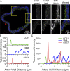
ASIC1 is expressed in mesenteric vascular smooth muscle and endothelial cells. (A) Cross-section of a mesenteric artery showing immunofluorescence of the endothelial marker, CD31 (green), ASIC1 (red), and smooth muscle α-actin (blue). Yellow rectangles represent zoomed-in images to the right. (B and C) The fluorescence intensity profile across the dotted white line shown in A for both individual (B) and merged (C) fluorescence intensity of CD31, SMA, and ASIC1.
Figure 2.
Figure 2.
ASIC1a contributes to ACh-induced vasodilation in mesenteric arteries. (A) Basal vessel wall intracellular [Ca2+] (F340/F380) in mesenteric arteries in the presence of vehicle or PcTX1 (20 nM). (B and C) Vasoconstriction (percent baseline inner diameter; B) and changes in vessel wall [Ca2+] (Δ F340/F380) in response to PE (10−8 to 10−5 M; C) in the presence of vehicle or PcTX1. (D) Representative traces showing changes in vessel inner diameter (µm) in response to increasing doses of ACh (10−9 to 10−5 M) in preconstricted mesenteric arteries. (E and F) The percent peak (E) and sustained (F) vasodilatory responses to ACh in the presence or absence of PcTX1. n = 5–6 animals (biological replicates)/group, analyzed by unpaired t tests, *P < 0.05 vs. vehicle. P values for data in E from 10−9 to 10−5 M ACh are 0.0158, 0.0384, 0.0017, 0.1907, 0.2096, 0.9620, 0.1264, 0.0415, and 0.0430. P values for data in F from 10−9 to 10−5 M ACh are 0.0269, 0.0661, 0.0031, 0.1524, 0.0884, 0.0151, 0.0008, 0.0014, and 0.0041.
Figure 3.
Figure 3.
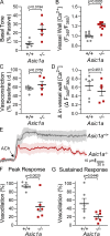
Augmented basal tone and blunted ACh-induced vasodilation in mesenteric arteries from Asic1a/ mice. (A and B) Basal tone (percent passive diameter; A) and basal vessel wall intracellular [Ca2+] (F340/F380; B) in mesenteric arteries from Asic1a+/+ or Asic1a/ mice. (C and D) Vasoconstriction (percent baseline inner diameter; C) and changes in the vessel wall [Ca2+] (Δ F340/F380; D) in response to Ang II (10−7 M). (E) Averaged traces showing changes in vessel inner diameter (µm) in response to ACh (10−6 M) in preconstricted mesenteric arteries from Asic1a+/+ or Asic1a/ mice. (F and G) Summary data showing percent peak (F) and sustained (G) vasodilatory responses to ACh. Biological replicates were analyzed by unpaired t tests.
Figure 4.
Figure 4.
ASIC1a contributes to ACh-induced Ca2+ entry in mesenteric endothelial tubes. (A) Endothelial tubes were freshly isolated and loaded with fura-2. (B) Averaged traces showing Mn2+-quenching of fura-2 fluorescence at 360 nm excitation. Experiments were conducted under basal conditions or following ACh (10−6 M) in the absence or presence of PcTX1 (20 nM). (C) Summary data for percent quenched fura-2 fluorescence by Mn2+ at 5 min. Dotted line represents mean basal values. Data represent biological replicates and were analyzed by two-way ANOVA. The two-way ANOVA revealed that there was a statistically significant interaction (P = 0.0003) between factors. Post-hoc pairwise comparisons were analyzed between individual groups using Tukey’s multiple comparisons test.
Figure 5.
Figure 5.
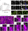
PKC activates ASIC1 in mesenteric endothelial tubes. (A) Representative immunofluorescence images in mesenteric endothelial tubes showing ASIC1a (red) and STIM-1 (green) distribution. Nuclei are stained with Sytox (blue). (B) Representative images showing lack of Duolink proximity ligation interaction between ASIC1–STIM1 (no observed red puncta). (C and D) Averaged traces (C) and summary data (D) showing Mn2+-quenching of fura-2 fluorescence (%) at 360 nm excitation. MnCl2 (500 µM) was added under vehicle conditions (0 Ca2+) or in the absence or presence of PcTX1 (20 nM) following incubation with CPA (10 µM) to deplete intracellular Ca2+ stores. Dotted line represents the absence of CPA (Veh). (E) Summary data showing Mn2+-quenching of fura-2 fluorescence (%) under basal conditions (dotted line) or following 5 min ACh (10−6 M) in the absence or presence of PLA2 inhibitor, MAF (5 µM), and/or PcTX1 (20 nM). Data in D and E represent biological replicates and were analyzed by one-way ANOVA. Post-hoc pairwise comparisons were analyzed between individual groups using Tukey’s multiple comparisons test. (F) Vasodilation responses to AA (10−8 to 3 × 10⁻⁵ M) following preconstriction with U-46619 in mesenteric arteries. (G) Summary data showing Mn2+-quenching of fura-2 fluorescence (%) following 10 min PMA (10−5 M) in the absence or presence of PcTX1 (20 nM). Biological replicates were analyzed by unpaired t tests.
Figure 6.
Figure 6.
ASIC1 contributes to endothelium-dependent, hyperpolarization-induced vasodilation, colocalizes with IKCa and SKCa channels, and localizes within myoendothelial junctions. (A) ACh (10−9–10−5 M) induced vasodilatory responses in mesenteric arteries, in the absence or presence of the NOS-specific inhibitor L-NNA (100 µM), the cyclooxygenase specific inhibitor indomethacin (Indo; 10 µM), and either PcTX1 (20 nM) or Tram-34 and Apamin (Tram/Ap; 1 µM and 100 nM, respectively). Data represent five animals (biological replicates)/group and were analyzed by two-way ANOVA. The two-way ANOVA revealed that there was a statistically significant interaction (P < 0.0001) between factors. Post hoc pairwise comparisons were analyzed between individual groups using Tukey’s multiple comparisons test. *P < 0.05 vehicle vs. L-NNA, Indo; #P < 0.05 L-NNA, Indo alone vs. PcTX1 or Tram/Ap. P values for data in A are shown in Table 2. (B and C) Immunofluorescence in mesenteric endothelial tubes showing ASIC1a (red) and (B) IKCa (green) or (C) SKCa (green) distribution. Nuclei are stained with Sytox (blue). (D) Representative images showing the Duolink proximity ligation interaction between ASIC1-IKCa, ASIC1-SKCa, or no primary antibodies as a negative control. (E) Autofluorescence (magenta) of the IEL and immunofluorescence of ASIC1 (green) in an intact, whole-mount mesenteric artery. Areas without magenta demonstrate fenestrations in IEL associated with myoendothelial junctions. Magnified images (a-f; yellow boxes) show the orthogonal view of the IEL layers (arrows), with the smooth muscle on the outside and endothelium between the IEL layers.
Figure 7.
Figure 7.
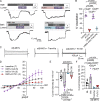
ASIC1 inhibition abolishes ACh-induced IKCa/SKCa currents. (A) Representative fluorescence and brightfield images showing identification of lectin-stain endothelial cells. (B–D) Representative K+ current traces and average current density (pA/pF) plotted as a function of voltage (mV) recorded in freshly dispersed mesenteric endothelial cells during baseline conditions (C) and following 1 µM ACh (D) in the presence of vehicle (black circles), PcTX1 (20 nM, red triangles), or Tram-34 (1 µM) and Apamin (100 nM; blue squares). Data represent biological replicates (noted in parentheses) and were analyzed by two-way ANOVA. The two-way ANOVA revealed that there was a statistically significant interaction (P < 0.0001) between factors. (E and F) Post-hoc pairwise comparisons were analyzed between individual groups using Tukey’s multiple comparisons test and the P values are given in Table 3 and summary data for −140 mV (E) and 100 mV (F). (G) Summary data showing Tram/Ap-sensitive and PcTX1-sensitive K+ currents under baseline conditions or following ACh.
Figure 8.
Figure 8.
Direct stimulation of ASIC1 causes vasodilation and activates IKCa/SKCa currents. (A and B) Representative trace showing changes in vessel inner diameter (µm; A) and summary data showing percent vasodilation in response to luminal perfusion of α/β-MitTx (200 nM bolus; B) in preconstricted mesenteric arteries in the presence of vehicle, PcTX1, LNNA-Indo, or LNNA-Indo-TRAM/Ap. Data represent biological replicates and were analyzed by one-way ANOVA. Post-hoc pairwise comparisons were analyzed between individual groups using Tukey’s multiple comparisons test. (C and D) Representative K+ current traces (C) and average current density (pA/pF; D) plotted as a function of voltage (mV) recorded in freshly dispersed mesenteric endothelial cells during baseline (from Fig. 7 B) and following activation of ASIC1 with α/β-MitTx (20 nM; purple diamond) in the absence or presence of PcTX1 (20 nM, red squares) or Tram-34 (1 µM) and Apamin (100 nM; blue triangles). Data represent biological replicates (noted in parentheses) and were analyzed by two-way ANOVA. The two-way ANOVA revealed that there was a statistically significant interaction (P < 0.0001) between factors. (E and F) Post-hoc pairwise comparisons were analyzed between individual groups using Tukey’s multiple comparisons test and the P values are given in Table 3 and in summary data for −140 mV (E) and 100 mV (F).
Figure 9.
Figure 9.
ASIC1a signaling in mesenteric endothelial cells. Left: In mesenteric endothelial cells, ASIC1a is activated by ACh. In the present study, we show this is likely due to the secondary activation via PKC, and not through endoplasmic reticulum (ER) store-depletion mechanisms involving STIM1 or AA. Right: Myoendothelial junction showing the presence of ASIC1a in the fenestrations of the IEL where it contributes to ACh-induced endothelium-dependent dilation through activation of IKCa/SKCa channels.

References

    1. Adapala, R.K., Talasila P.K., Bratz I.N., Zhang D.X., Suzuki M., Meszaros J.G., and Thodeti C.K.. 2011. PKCα mediates acetylcholine-induced activation of TRPV4-dependent calcium influx in endothelial cells. Am. J. Physiol. Heart Circ. Physiol. 301:H757–H765. 10.1152/ajpheart.00142.2011 - DOI - PMC - PubMed
    1. Akanji, O., Weinzierl N., Schubert R., and Schilling L.. 2019. Acid sensing ion channels in rat cerebral arteries: Probing the expression pattern and vasomotor activity. Life Sci. 227:193–200. 10.1016/j.lfs.2019.04.054 - DOI - PubMed
    1. Baron, A., Deval E., Salinas M., Lingueglia E., Voilley N., and Lazdunski M.. 2002. Protein kinase C stimulates the acid-sensing ion channel ASIC2a via the PDZ domain-containing protein PICK1. J. Biol. Chem. 277:50463–50468. 10.1074/jbc.M208848200 - DOI - PubMed
    1. Bashari, E., Qadri Y.J., Zhou Z.-H., Kapoor N., Anderson S.J., Meltzer R.H., Fuller C.M., and Benos D.J.. 2009. Two PKC consensus sites on human acid-sensing ion channel 1b differentially regulate its function. Am. J. Physiol. Cell Physiol. 296:C372–C384. 10.1152/ajpcell.00200.2008 - DOI - PMC - PubMed
    1. Benarroch, E.E. 2014. Acid-sensing cation channels: Structure, function, and pathophysiologic implications. Neurology. 82:628–635. 10.1212/WNL.0000000000000134 - DOI - PubMed

Publication types

Substances