. 2022 Dec 9;8(1):219.
doi: 10.1186/s40792-022-01576-6.

Laparoscopic resection of an intra-abdominal esophageal duplication cyst in the ileum: a case report

Affiliations

Laparoscopic resection of an intra-abdominal esophageal duplication cyst in the ileum: a case report

Shosaburo Oyama et al. Surg Case Rep. .

Abstract

Background: Esophageal duplication cyst (EDC) is a type of gastrointestinal duplication cyst that involves congenital malformations of the gastrointestinal tract. EDCs are frequently found in the mediastinum and thoracoabdominal region, but rarely occur in the abdominal cavity. However, intra-abdominal EDCs are frequently found in the upper abdomen near the abdominal esophagus. Here, we report, for the first time, a case of intra-abdominal EDC that occurred in the ileum.

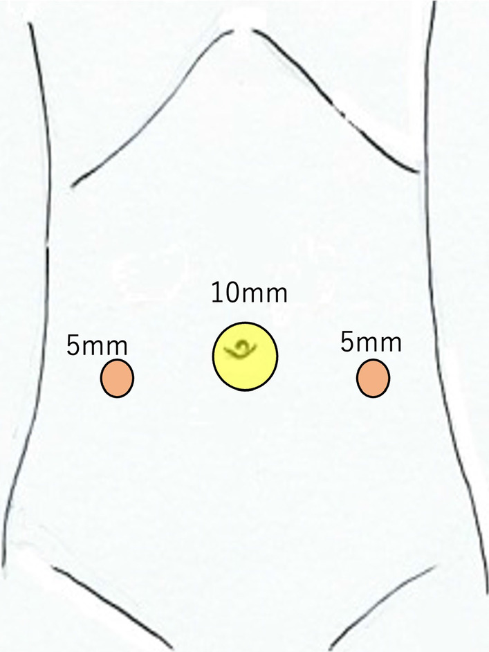
Case presentation: A 14-year-old female patient presented to our hospital with complaints of epigastric pain and vomiting. Abdominal computed tomography (CT) revealed a cystic tumor in the pelvis, suspected of ovarian origin. She was admitted to our gynecology department and underwent emergency surgery. The laparoscopic examination revealed that both ovaries were intact and that a primary tumor had developed from the ileal mesentery. Since the patient's condition was not urgent at the time of the gynecological surgery, the procedure was completed by only performing exploratory laparotomy; the patient was admitted to our department after the surgery. Pelvic magnetic resonance imaging performed on the next day revealed a cystic mass measuring 90 × 65 mm with a smooth margin and homogeneous signal intensity, arising posterior to the uterus. The mass was suspected as an intestinal duplication cyst. On another day, after the examinations were completed, we resected the portion of the small intestine containing the tumor by laparoscopy. The patient had a successful postoperative course and was discharged on the 5th postoperative day. Histological examination showed that the cyst was lined by stratified squamous epithelium, contained esophageal glands, and had a two-layer muscularis propria. Therefore, a diagnosis of intra-abdominal EDC was performed.

Conclusions: An intra-abdominal EDC cyst is relatively rare; this is the first case reported at the distal ileum.

Keywords: Ileum; Intra-abdominal esophageal duplication cyst; Laparoscopic surgery.


Conflict of interest statement

The authors declare that they have no competing interest.

Figures

Fig. 1
Fig. 1
Preoperative pelvic magnetic resonance imaging. Preoperative images show a tumor (yellow arrow) that was a cystic mass measuring 90 × 65 mm with a smooth margin and homogeneous signal intensity on the right dorsal side of the uterus (ad). Coronal image (c), sagittal image (d). T1-weighted images present slightly higher signals than those of water, and T2-weighted images show low signals. No apparent communication was found between the cyst and the intestinal tract
Fig. 2
Fig. 2
Port site placement. The image shows the three ports of laparoscopic surgery. The umbilical port is a 12-mm camera port (yellow circle), and the bilateral abdomen port is a 5-mm working port for handling the forceps (red circle)
Fig. 3
Fig. 3
Intraoperative laparoscopic view. The image shows a tumor as a lesion that fits into the pelvis (a). The lesion has good mobility without adhesions in the pelvis and can exit the body cavity from the umbilical region (b, c). The cystic tumor was located on the mesenteric side, approximately 40 cm proximal to the distal ileum. The boundary with the adjacent intestinal tract is unclear in the intraoperative findings, and partial resection of the small intestine containing the tumor was performed
Fig. 4
Fig. 4
Extracted specimen. The image shows the specimen at the ileal lumen (a). No change in the lumen and no apparent communication were observed with the cyst. The image shows the lumen of the cyst (b)
Fig. 5
Fig. 5
Histopathological findings. The cystic epithelium appears as stratified squamous epithelium (hematoxylin–eosin [HE], ×100) (a). The esophageal glands are observed in the submucosa, which has mucous glands and ducts (HE, ×100) (b). The muscular layer has a two-layer structure with inner circular and outer longitudinal muscle layers (c). In addition, nerves and ganglion cells (Auerbach plexus, yellow arrow) are found between the two layers (HE, ×100)

Similar articles

Cited by

References

    1. Mori M, Shuto K, Hirano A, Kosugi C, Narushima K, Koda K. Laparoscopic surgery for an esophageal duplication cyst using a near-infrared indocyanine green fluorescence system: a case report. Asian J Endosc Surg. 2020;13:211–214. doi: 10.1111/ases.12729. - DOI - PMC - PubMed
    1. Pujar VC, Kurbet S, Kaltari DK. Laparoscopic excision of intra-abdominal oesophageal duplication cyst in a child. J Minim Access Surg. 2013;9:34–36. doi: 10.4103/0972-9941.107137. - DOI - PMC - PubMed
    1. Harvell JD, Macho JR, Klein HZ. Isolated intra-abdominal esophageal cyst. Case report and review of the literature. Am J Surg Pathol. 1996;20:476–479. doi: 10.1097/00000478-199604000-00011. - DOI - PubMed
    1. Martin ND, Kim JC, Verma SK, Rubin R, Mitchell DG, Bergin D, et al. Intra-abdominal esophageal duplication cysts: a review. J Gastrointest Surg. 2007;11:773–777. doi: 10.1007/s11605-007-0108-0. - DOI - PubMed
    1. Bromberg SH, Camilo Neto C, Borges AF, Franco MI, França LC, Yamaguchi N. Pancreatic heterotopias: clinicopathological analysis of 18 patients. Rev Col Bras Cir. 2010;37:413–419. doi: 10.1590/s0100-69912010000600007. - DOI - PubMed

LinkOut - more resources