. 2022 Dec 9;8(49):eadd4187.
doi: 10.1126/sciadv.add4187. Epub 2022 Dec 9.

Autoinhibition and activation mechanisms revealed by the triangular-shaped structure of myosin Va

Affiliations

Autoinhibition and activation mechanisms revealed by the triangular-shaped structure of myosin Va

Fengfeng Niu et al. Sci Adv. .

Abstract

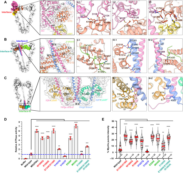
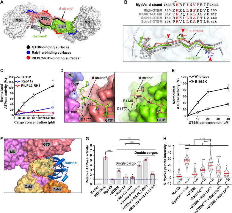
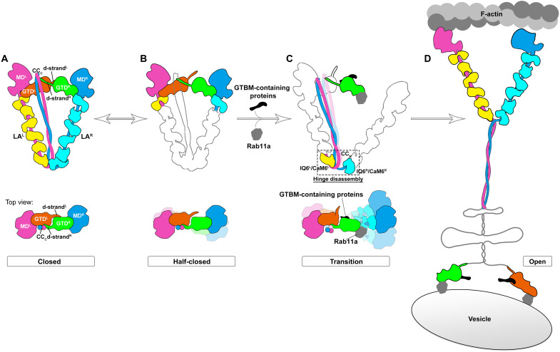
As the prototype of unconventional myosin motor family, myosin Va (MyoVa) transport cellular cargos along actin filaments in diverse cellular processes. The off-duty MyoVa adopts a closed and autoinhibited state, which can be relieved by cargo binding. The molecular mechanisms governing the autoinhibition and activation of MyoVa remain unclear. Here, we report the cryo-electron microscopy structure of the two full-length, closed MyoVa heavy chains in complex with 12 calmodulin light chains at 4.78-Å resolution. The MyoVa adopts a triangular structure with multiple intra- and interpolypeptide chain interactions in establishing the closed state with cargo binding and adenosine triphosphatase activity inhibited. Structural, biochemical, and cellular analyses uncover an asymmetric autoinhibition mechanism, in which the cargo-binding sites in the two MyoVa heavy chains are differently protected. Thus, specific and efficient MyoVa activation requires coincident binding of multiple cargo adaptors, revealing an intricate and elegant activity regulation of the motor in response to cargos.


Figures

Fig. 1.
Fig. 1.. Overall structure of the closed MyoVa-CaM4M complex.
(A) Schematic representation of the MyoVa-CaM complex. The domains of MyoVa and six CaM molecules are indicated. (B) Cryo-EM map of the MyoVa-CaM4M complex in the closed state, colored by different molecules. Two GTDs from the two MyoVa heavy chains are independently colored because of the undefined connection to the CC. The triangular shape is highlighted with the indicated lengths of three sides. (C to E) Top and bottom views (C and D) and two side views (E) of the cryo-EM map show the twisted triangular shape of the MyoVa-CaM4M complex. The density of two d-strands for GTD dimerization and two hinge strands for the CC reverse is labeled. The GTD plane, hinge plane, the twofold symmetric axis, and two long sides with different curvatures are indicated. (F) Atomic model of the MyoVa-CaM4M complex. All molecules or domains are colored with the same code as shown in (B). SD-I/SD-II of the GTDs, the two helices of the CC, and the N-/C-lobe of CaM are indicated. (G) Structural comparisons of the head/tail, LA, and hinge/CC regions between MyoVaL and MyoVaR. The two LAs and the hinge and CC regions are compared by aligning the IQ1/CaM1 and the IQ6/CaM6, respectively.
Fig. 2.
Fig. 2.. Comparison of our MyoVa-CaM4M complex structure to the previous model.
(A) Structural comparison of our model and the reported model [Protein Data Bank (PDB) ID: 2DFS] from a previous low-resolution map (~24 Å at FSC = 0.5). The GTD dimer in our model is indicated. The comparison of the MD, CC, and hinge regions are shown as zoom-in views in (B), (C), and (D), respectively. (B) Structural comparison of the MDL regions in the two models. The GTD in our closed structure is showed with a transparent surface. D134 in the GTD-binding interface and the loop-1 close to the active site of MDs are highlighted, and their rotations between the two models are indicated by white arrows. (C) Structural comparison of the CC regions in the two models. The two ends of the CC that were not built in the previous model are indicated by two red dashed circles, respectively. (D) Structural comparison of the hinges in the two models. MyoVa heavy chain and CaM6 are highlighted in the left and right panels for the comparison, respectively. The N-lobe and C-lobe of CaM6 are indicated.
Fig. 3.
Fig. 3.. The interactions in the formation of the closed MyoVa-CaM4M complex.
(A to C) The four interfaces in the MyoVa-CaM4M complex, including the interface-I between MDL/CaM1L and GTDL for the head/GTD interaction (A), the interface-II between GTDL and d-strandR for the GTD dimerization (B), the interface-III between IQ6/CaM6 and CCN for the hinge formation (C), and the interface-IV between GTDL and CCC (B). The molecular details of these interfaces are presented in zoom-in views with the interacting residues shown in stick mode. Hydrogen bonds and salt bridges are indicated as dashed lines. V1437 that was mutated to phenylalanine for structure determination is labeled with an asterisk. (D) The ATPase activity analysis of the wild-type and mutated MyoVa in complex with CaM. The MD(2IQs) was used to indicate the fully activated MD. All measured activity values were normalized to that of the wild-type MyoVa for comparison. All the reactions were experimentally repeated three times in this and the following measurements of ATPase activity. (E) Statistical analysis of the puncta formation of MyoVa or its mutants in the HeLa cells. The representative imaging data are shown in fig. S8C. Cell number for each condition investigated is indicated. The mutations locating in interfaces I to IV are respectively labeled with different color in (D) and (E), corresponding to the interfaces I to IV indications in (A) to (C). ns, not significant.
Fig. 4.
Fig. 4.. Structural analysis of MD and its inhibition in the closed state.
(A) GTD-bound MD structure in the closed MyoVa. The four subdomains, N-terminal, U50, L50, and converter in MD are indicated, which regulates the coupled conformational change of the actin-binding site, active site, and LA. In the active site, P-loop, switch-I, and switch-II, essential for ATP binding and hydrolysis, are colored in red, blue, and green, respectively. The nucleotide-binding site is marked by a star. The helix in CaM1 that binds to the GTD is indicated by a red arrowhead. (B) Structural comparisons of MD and LA in the closed state (GTD-bound) to those in rigor (PDB ID: 7PLT)/rigor-like (1OE9), post-rigor transition (7PMD), post-rigor (1W7J), pre-powerstroke (4ZG4), and weak/strong ADP-binding (1W7I and 7PM5) states within the ATPase cycle by aligning their N-terminal subdomains. The structural model of the pre-powerstroke state is generated by adding IQ1/CaM1 to the MyoVc-MD structure with the pre-powerstroke conformation. The LA rotation angles between the closed state and the other states are indicated. (C) Structural comparisons of active sites in the closed state and the states of the ATPase cycle showing the conformational changes of switch-I, switch-II, and P-loop. The bound nucleotides are shown in sticks. (D) Structural comparisons of actin-binding surfaces in the closed state, the different states in the ATPase cycle by aligning their subdomains of the U50 of the MD, and the conformational changes of the subdomain of the L50 are indicated.
Fig. 5.
Fig. 5.. Cargo-mediated MyoVa activation.
(A) Three cargo-binding surfaces on the GTD that are blocked in the closed state. (B) Sequence and structural alignments of the d-strand in MyoVa and GTBMs in Mlph (PDB ID: 4KP3), MICAL1 (6KU0), and Spires (5JCY) by aligning the GTDs. The three key residues for GTD binding are highlighted. (C) ATPase activity of MyoVa (0.1 μM) measured with the different concentrations of indicated proteins. The activity was normalized to the conditions containing MyoVa and MD(2IQs) as 0 and 100%, respectively. Mlph-GTBM was used in this and the following ATPase assays. (D) Sheltering effect of CCC on the GTDL/d-strandR, but not GTDR/d-strandL, interaction. (E) ATPase activities of MyoVa and MyoVaE1089K (0.1 μM) in the presence of Mlph-GTBM. (F) Head/GTD interaction is disrupted by the binding of Rab11a to the GTD. The Rab11a-bound GTD (PDB ID: 5JCZ) is superimposed with that in the closed state. Clashes between the CaM1 and Rab11a are indicated by a dashed circle. (G) ATPase activity of MyoVa in the presence of individual cargo adaptors or their combinations. Concentrations of MyoVa and each cargo are of 0.1 and 40 μM, respectively. The ATPase activities were normalized with the same method used in Fig. 3D. (H) Statistical analysis of the punctate MyoVa in the HeLa cells coexpressing the wild-type MyoVa with either Spire1-GTBM or Rab11aQ70L (a constitutively active form) or both Spire-GTBM and Rab11aQ70L. The GTD binding-deficient mutants, Spire1-GTBML454Q and Rab11aF48Q, were analyzed correspondingly. The indicated number of cells was selected for analysis in each condition.
Fig. 6.
Fig. 6.. A proposed activation model of MyoVa.
(A) Closed state of MyoVa. The schematic drawing of this conformation is derived from the closed MyoVa-CaM4M structure. (B) Half-closed state of MyoVa. The d-strandL temporally dissociates from the GTDR, leading to the dynamic conformation of the short side in the triangular-shaped structure. (C) “Transition” state of MyoVa. In the presence of both a GTBM-containing protein and Rab11a, the half-closed conformation undergoes a large motion of both the MDR and the LAR, resulting in the disassembly of the hinge structure. Notably, only one cargo binding of either GTBM-containing protein or Rab11a is not sufficient to effectively activate MyoVa. (D) Open state of MyoVa. The unstable transition conformation quickly shifts to the open conformation, allowing the GTDs to tightly associate with cargos and the MDs to hydrolyze ATP for the walking.

References

    1. M. A. Hartman, D. Finan, S. Sivaramakrishnan, J. A. Spudich,Principles of unconventional myosin function and targeting. Annu. Rev. Cell Dev. Biol. 27,133–155 (2011). - PMC - PubMed
    1. N. Hirokawa, R. Takemura,Molecular motors and mechanisms of directional transport in neurons. Nat. Rev. Neurosci. 6,201–214 (2005). - PubMed
    1. N. Hirokawa, S. Niwa, Y. Tanaka,Molecular motors in neurons: Transport mechanisms and roles in brain function, development, and disease. Neuron 68,610–638 (2010). - PubMed
    1. M. Kneussel, W. Wagner,Myosin motors at neuronal synapses: Drivers of membrane transport and actin dynamics. Nat. Rev. Neurosci. 14,233–247 (2013). - PubMed
    1. R. D. Vale,The molecular motor toolbox for intracellular transport. Cell 112,467–480 (2003). - PubMed