. 2023 Feb 16;141(7):725-742.
doi: 10.1182/blood.2022015917.

The histone methyltransferase MLL1/KMT2A in monocytes drives coronavirus-associated coagulopathy and inflammation

Affiliations

The histone methyltransferase MLL1/KMT2A in monocytes drives coronavirus-associated coagulopathy and inflammation

Sriganesh B Sharma et al. Blood. .

Abstract

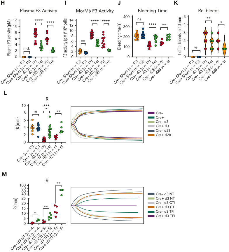
Coronavirus-associated coagulopathy (CAC) is a morbid and lethal sequela of severe acute respiratory syndrome coronavirus 2 (SARS-CoV-2) infection. CAC results from a perturbed balance between coagulation and fibrinolysis and occurs in conjunction with exaggerated activation of monocytes/macrophages (MO/Mφs), and the mechanisms that collectively govern this phenotype seen in CAC remain unclear. Here, using experimental models that use the murine betacoronavirus MHVA59, a well-established model of SARS-CoV-2 infection, we identify that the histone methyltransferase mixed lineage leukemia 1 (MLL1/KMT2A) is an important regulator of MO/Mφ expression of procoagulant and profibrinolytic factors such as tissue factor (F3; TF), urokinase (PLAU), and urokinase receptor (PLAUR) (herein, "coagulopathy-related factors") in noninfected and infected cells. We show that MLL1 concurrently promotes the expression of the proinflammatory cytokines while suppressing the expression of interferon alfa (IFN-α), a well-known inducer of TF and PLAUR. Using in vitro models, we identify MLL1-dependent NF-κB/RelA-mediated transcription of these coagulation-related factors and identify a context-dependent, MLL1-independent role for RelA in the expression of these factors in vivo. As functional correlates for these findings, we demonstrate that the inflammatory, procoagulant, and profibrinolytic phenotypes seen in vivo after coronavirus infection were MLL1-dependent despite blunted Ifna induction in MO/Mφs. Finally, in an analysis of SARS-CoV-2 positive human samples, we identify differential upregulation of MLL1 and coagulopathy-related factor expression and activity in CD14+ MO/Mφs relative to noninfected and healthy controls. We also observed elevated plasma PLAU and TF activity in COVID-positive samples. Collectively, these findings highlight an important role for MO/Mφ MLL1 in promoting CAC and inflammation.


Conflict of interest statement

Conflict-of-interest disclosure: M.H. is a consultant and equity holder for Veralox Therapeutics and a consultant for Cereno Scientific, which has an option to license platelet inhibitory compounds from the University of Michigan. The remaining authors declare no competing financial interests.

Figures

None
Graphical abstract
Figure 1.
Figure 1.
Coronavirus infection of MO/Mφs induces expression of MLL1, coagulopathy-related factors, and inflammatory cytokines. BMDMs were harvested from C57Bl6/J mice (wild-type-BMDMs) and were infected with 1 MOI of the murine coronavirus MHVA59 for the indicated times, and mRNA (A) and protein levels (B; representative blot shown [β-actin served as loading control]) of Kmt2a/MLL1 were assayed by qRT-PCR and immunoblotting, respectively. (C) mRNA levels of factors important in CAC (Plau, Plaur, and F3) were measured in infected BMDMs. (D) mRNA levels of proinflammatory cytokines identified in the inflammatory signature resulting from acute SARS-CoV-2 (IL-6 and TNFα) and the MLL1-regulated cytokine IL-1β were measured in infected BMDMs. (E-F) Protein levels of coagulopathy-related factors (E) and proinflammatory cytokines (F) were assayed by ELISA. Bar graphs represent mean values from at least n = 5 independent experiments assayed in triplicate, and individual data points represent independent experiments. Errors bars represent SE. Statistical testing was performed using Kruskal-Wallis tests with corrections for multiple comparisons. ∗P < .05; ∗∗P < .01; ∗∗∗P < .001; ∗∗∗∗P < .0001. ELISA, enzyme-linked immunosorbent assay; MOI, multiplicity of infection; qRT, quantitative reverse transcription; SE, standard error.
Figure 2.
Figure 2.
MLL1 regulates the basal expression of coagulopathy-related factors and proinflammatory cytokines in BMDMs. BMDMs were harvested from mice carrying a myeloid-specific deletion of MLL1 (Kmt2afl/fl Lyz2Cre+/−; denoted Cre+) and littermate controls (Kmt2afl/fl Lyz2Cre−/−; denoted Cre−). (A) mRNA levels of Kmt2a were assayed in n = 8 Cre+ and Cre− animals analyzed in triplicate. (B) Protein levels of MLL1 were assayed in BMDMs from n = 4 Cre+ and Cre− mice by immunoblotting (representative blot shown [β-actin served as loading control]). (C-D) mRNA levels of coagulopathy-related factors (C) and proinflammatory cytokines (D) were assayed. (E-F) Protein levels of coagulopathy-related factors (E) and inflammatory cytokines (F) were measured by ELISA. (G) ChIP assays were performed using antibodies specific to either H3K4me3 or nontargeting species-specific IgG. Bars graphs represent mean values from at least n = 4 independent experiments with individual data points representing independent experiments. For ChIP experiments, bar graphs represent mean ChIP intensity relative to IgG derived from n = 8 samples performed in triplicate. Statistical analysis of pairwise comparisons was performed using Mann-Whitney tests. Error bars represent SE. ∗P < .05; ∗∗P < .01; ∗∗∗P < .001; ∗∗∗∗P < .0001. ChIP, chromatin immunoprecipitation; ELISA, enzyme-linked immunosorbent assay; IgG, immunoglobulin G; SE, standard error.
Figure 3.
Figure 3.
Loss of MLL1 attenuates coronavirus and proinflammatory cytokine-mediated induction of coagulopathy-related factors and proinflammatory cytokines in BMDMs. BMDMs were harvested from mice carrying a myeloid-specific deletion of MLL1 (Kmt2afl/fl Lyz2Cre+/−; denoted Cre+) and littermate controls (Kmt2afl/fl Lyz2Cre−/−; denoted Cre−). Cells were either stimulated with TNFα (5 ng/mL), IL-1β (5 ng/mL), or IL-6 (5 ng/mL) or infected with 1 MOI of MHVA59 for 24 hours. (A-B) mRNA levels of coagulopathy-associated factors and inflammatory cytokines were assayed in stimulated/infected cells by qRT-PCR. (C-D) Protein levels of coagulopathy-related factors (C) and inflammatory cytokines (D) were measured by ELISA. Crossed circles indicate cytokines, which was not measured in each assay as the particular cytokine was used for stimulation. (E) ChIP assays were performed using antibodies specific for H3K4me3 or with nontargeting species-specific IgG antibodies. ChIP intensity (relative to IgG) at the indicated promoters is shown. For qRT-PCR and ELISA experiments, graphs feature results from n = 10 animals assayed in triplicate. For ChIP experiments, bar graphs represent mean ChIP intensity relative to IgG from n = 8 animals assayed in triplicate. Error bars represent SE. For clarity of figure, graphical representation of statistical significance is not shown. Statistical analysis of data sets was performed using Mann-Whitney U tests and pairwise comparisons between Cre− and Cre+ cells were found to meet criteria for statistical significance (P < .05) except where indicated. ChIP, chromatin immunoprecipitation; ELISA, enzyme-linked immunosorbent assay; IgG, immunoglobulin G; MOI, multiplicity of infection; qRT, quantitative reverse transcription; SE, standard error.
Figure 3.
Figure 3.
Loss of MLL1 attenuates coronavirus and proinflammatory cytokine-mediated induction of coagulopathy-related factors and proinflammatory cytokines in BMDMs. BMDMs were harvested from mice carrying a myeloid-specific deletion of MLL1 (Kmt2afl/fl Lyz2Cre+/−; denoted Cre+) and littermate controls (Kmt2afl/fl Lyz2Cre−/−; denoted Cre−). Cells were either stimulated with TNFα (5 ng/mL), IL-1β (5 ng/mL), or IL-6 (5 ng/mL) or infected with 1 MOI of MHVA59 for 24 hours. (A-B) mRNA levels of coagulopathy-associated factors and inflammatory cytokines were assayed in stimulated/infected cells by qRT-PCR. (C-D) Protein levels of coagulopathy-related factors (C) and inflammatory cytokines (D) were measured by ELISA. Crossed circles indicate cytokines, which was not measured in each assay as the particular cytokine was used for stimulation. (E) ChIP assays were performed using antibodies specific for H3K4me3 or with nontargeting species-specific IgG antibodies. ChIP intensity (relative to IgG) at the indicated promoters is shown. For qRT-PCR and ELISA experiments, graphs feature results from n = 10 animals assayed in triplicate. For ChIP experiments, bar graphs represent mean ChIP intensity relative to IgG from n = 8 animals assayed in triplicate. Error bars represent SE. For clarity of figure, graphical representation of statistical significance is not shown. Statistical analysis of data sets was performed using Mann-Whitney U tests and pairwise comparisons between Cre− and Cre+ cells were found to meet criteria for statistical significance (P < .05) except where indicated. ChIP, chromatin immunoprecipitation; ELISA, enzyme-linked immunosorbent assay; IgG, immunoglobulin G; MOI, multiplicity of infection; qRT, quantitative reverse transcription; SE, standard error.
Figure 4.
Figure 4.
MLL1 is required for RelA-dependent transcription of coagulopathy-associated factors. (A) Rela mRNA levels were assayed in BMDMs harvested from MLL1 knockout mice (Cre+; n = 4) and littermate controls (Cre−; n = 4). (B) RelA protein levels are assayed in these cells by immunoblot (β-actin served as loading control). (C) The protein expression of MLL1 (MLL1C = C-terminal epitope), phospho-RelA (Ser536), and RelA was assayed in RAW264.7 cells that were transfected with either of 2 distinct siRNAs targeting MLL1 (denoted −1 and −2) or a nontargeting control (-Ctl). (β-actin served as loading control) (D) mRNA expression of Rela and Kmt2a was assayed in BMDMs transfected with siRNAs targeting Rela (smartpool; denoted siRelA) or a nontargeting control (siCtl). Results are representative of n = 4 independent experiments assayed in triplicate. (E) Protein levels of MLL1 and RelA were assayed, and representative immunoblot is shown. (F) BMDMs from littermate control mice (n = 8, assayed in triplicate) were transfected with the siRNAs and were infected with 1 MOI of MHVA59 for 24 hours, and mRNA levels of Kmt2a were measured. (G) Cre− and Cre+ BMDMs (n = 7 per group; assayed in triplicate) were transfected as indicated and infected with 1 MOI of MHVA59 for 24 hours and mRNA levels of Rela were assayed in these cells. (H) Cre− and Cre+ BMDMs (n = 7 per group; assayed in triplicate) were infected with MHVA59 for the indicated duration and ChIP assays were performed with antibodies specific to RelA or IgG. (I) mRNA levels of coagulopathy-associated factors were assayed in the indicated transfected cells (n = 7; performed in triplicate). (J) Protein levels of these coagulopathy-related factors were measured by ELISA (n = 7; performed in triplicate). (K) RelA ChIP assays were performed on candidate promoters and ChIP intensity relative to IgG was measured at the indicated promoters (n = 7 per group; assayed in triplicate). Bar graphs represent mean values and error bars represent SE. Statistical analysis was performed by either Mann-Whitney U test or Kruskal-Wallis test with corrections for multiple comparisons. Error bars represent SE. ∗P < .05; ∗∗P < .01; ∗∗∗P < .001; ∗∗∗∗P < .0001. ChIP, chromatin immunoprecipitation; ELISA, enzyme-linked immunosorbent assay; IgG, immunoglobulin G; MOI, multiplicity of infection; SE, standard error.
Figure 4.
Figure 4.
MLL1 is required for RelA-dependent transcription of coagulopathy-associated factors. (A) Rela mRNA levels were assayed in BMDMs harvested from MLL1 knockout mice (Cre+; n = 4) and littermate controls (Cre−; n = 4). (B) RelA protein levels are assayed in these cells by immunoblot (β-actin served as loading control). (C) The protein expression of MLL1 (MLL1C = C-terminal epitope), phospho-RelA (Ser536), and RelA was assayed in RAW264.7 cells that were transfected with either of 2 distinct siRNAs targeting MLL1 (denoted −1 and −2) or a nontargeting control (-Ctl). (β-actin served as loading control) (D) mRNA expression of Rela and Kmt2a was assayed in BMDMs transfected with siRNAs targeting Rela (smartpool; denoted siRelA) or a nontargeting control (siCtl). Results are representative of n = 4 independent experiments assayed in triplicate. (E) Protein levels of MLL1 and RelA were assayed, and representative immunoblot is shown. (F) BMDMs from littermate control mice (n = 8, assayed in triplicate) were transfected with the siRNAs and were infected with 1 MOI of MHVA59 for 24 hours, and mRNA levels of Kmt2a were measured. (G) Cre− and Cre+ BMDMs (n = 7 per group; assayed in triplicate) were transfected as indicated and infected with 1 MOI of MHVA59 for 24 hours and mRNA levels of Rela were assayed in these cells. (H) Cre− and Cre+ BMDMs (n = 7 per group; assayed in triplicate) were infected with MHVA59 for the indicated duration and ChIP assays were performed with antibodies specific to RelA or IgG. (I) mRNA levels of coagulopathy-associated factors were assayed in the indicated transfected cells (n = 7; performed in triplicate). (J) Protein levels of these coagulopathy-related factors were measured by ELISA (n = 7; performed in triplicate). (K) RelA ChIP assays were performed on candidate promoters and ChIP intensity relative to IgG was measured at the indicated promoters (n = 7 per group; assayed in triplicate). Bar graphs represent mean values and error bars represent SE. Statistical analysis was performed by either Mann-Whitney U test or Kruskal-Wallis test with corrections for multiple comparisons. Error bars represent SE. ∗P < .05; ∗∗P < .01; ∗∗∗P < .001; ∗∗∗∗P < .0001. ChIP, chromatin immunoprecipitation; ELISA, enzyme-linked immunosorbent assay; IgG, immunoglobulin G; MOI, multiplicity of infection; SE, standard error.
Figure 5.
Figure 5.
Coronavirus induces the expression of MLL1 and its associated factors in MO/Mφs in vivo and promotes a prothrombotic and profibrinolytic phenotype. C57BL6/J mice underwent intranasal inoculation of 2 × 105 plaque-forming units (pfu) MHVA59 (postinfection day 3 mice [d3; n = 8]; postinfection day 28 mice [d28; n = 9]) or PBS (denoted as sham; n = 8). Mice were sacrificed at the indicated time points and plasma and splenic MO/Mφs (a surrogate for circulating MO/Mφs) were harvested. (A) Kmt2a mRNA levels were assayed by qRT-PCR. (B-C) The mRNA levels and protein levels of coagulopathy-associated factors in splenic MO/Mφs were measured by qRT-PCR and ELISA, respectively. (D) H3K4me3 abundance at the indicated promoters was assayed by ChIP assay. (E-F) Circulating levels of PLAU and PLAUR were measured by ELISA. (G) Plasma TF protein levels was measured by ELISA. (H) Plasma PLAU activity levels were measured using a colorimetric assay in which absorbance (A405) correlates with enzyme activity level through the cleavage of a plasmin (activated by PLAU) substrate that liberates p-nitroaniline. (I) Plasma TF activity was measured using a colorimetric assay in which the activation of factor X (FXa) by TF and factor VII (TF/FVIIa) and its cleavage of a FXa-specific substrate liberates p-nitroaniline. (J) The TF activity of lysed harvested splenic MO/Mφs was measured. (K) Tail vein bleeding time was measured in infected and sham mice. (L) The number of rebleeding events during tail vein bleeding time assays was tallied. (M) Whole blood was collected from infected and sham mice by inferior vena cava puncture and anticoagulated with 3.2% sodium citrate at a ratio of 9:1 (blood to citrate). TEG was performed and R time (time to formation of clot of 2 mm thickness) was measured (left panel). Representative TEGs are presented in the right panel. (N) To determine the role of TF in hypercoagulability as assayed by a shortened R time as measured by TEG after coronavirus infection, citrated whole blood samples from either sham or infected (d3) mice was treated with either corn trypsin inhibitor (CTI; 25 μg/mL final concentration) or a mouse specific anti-TF neutralizing antibody (TFI; clone 1H1 [Genentech]; 50 μg/mL final concentration) and the resultant viscoelastic properties were analyzed using TEG. Samples that were not subjected to treatment (NT) served as controls. Bar graphs represent mean values. qRT-PCR, ELISA, and ChIP data represent experiments performed in triplicate. Error bars represent SE. Statistical analysis of data sets was performed by either Mann-Whitney U test or Kruskal-Wallis test with corrections for multiple comparisons. ∗P < .05; ∗∗P < .01; ∗∗∗P < .001; ∗∗∗∗P < .0001. ChIP, chromatin immunoprecipitation; ELISA, enzyme-linked immunosorbent assay; PBS, phosphate-buffered saline; qRT, quantitative reverse transcription; SE, standard error
Figure 6.
Figure 6.
Coronavirus-mediated induction of coagulopathy-related factors and the resultant coagulopathy is dependent on myeloid-specific expression of MLL1. Mice harboring a myeloid-specific MLL1 knockout (denoted Cre+; n indicated per group) and littermate controls (Cre−) underwent intranasal inoculation of 2 × 105 plaque-forming units (pfu) of MHVA59. Mice were sacrificed at the indicated time points and plasma and splenic MO/Mφs (a surrogate for circulating MO/Mφs) were harvested. (A-B) The mRNA and protein levels of coagulation associated factors were assayed by qRT-PCR and ELISA in splenic MO/Mφs, respectively. (C) H3K4me3 abundance at the indicated promoters was measured by ChIP assay. (D-E) Circulating levels of PLAU (D) and soluble PLAUR (E) were measured by ELISA. (F) Plasma PLAU activity levels were measured using a colorimetric assay in which absorbance (A405) correlates with enzyme activity level through the cleavage of a plasmin (activated by PLAU) substrate that liberates p-nitroaniline. (G) Plasma TF protein levels were measured by ELISA. (H) Plasma TF activity was measured using a colorimetric assay in which the activation of factor X (FXa) by TF and factor VII (TF/FVIIa) and its cleavage of a FXa-specific substrate liberates p-nitroaniline. (I) The TF activity of lysed harvested splenic MO/Mφs was measured. (J) Tail vein bleeding time was measured in infected and sham mice. (K) The number of rebleeding events during tail vein bleeding time assays was tallied. (L) Whole blood was collected from infected and sham mice by inferior vena cava puncture and anticoagulated with 3.2% sodium citrate at a ratio of 9:1 (blood to citrate). TEG was performed and R time (time to formation of clot of 2 mm thickness) was measured (left panel). Representative TEG is presented in the right panel. (M) To determine the role of TF in hypercoagulability as assayed by a shortened R time as measured by TEG after coronavirus infection, citrated whole blood samples from either infected (d3) Cre− or Cre+ mice was treated with either corn trypsin inhibitor (CTI; 25 μg/mL final concentration) or a mouse-specific anti-TF neutralizing antibody (TFI; clone 1H1 [Genentech]; 50 μg/mL final concentration) and the resultant viscoelastic properties were analyzed using TEG. Samples which were not subjected to treatment (NT) served as controls. Representative TEG is presented in the right panel. Bar graphs represent mean values and number of independent experiments per panel is as indicated. qRT-PCR, ELISA, and ChIP experiments were performed in triplicate. Error bars represent SE. Statistical comparisons were performed by either Mann-Whitney U test or Kruskal-Wallis test with corrections for multiple comparisons. ∗P < .05; ∗∗P < .01; ∗∗∗P < .001; ∗∗∗∗P < .0001. ChIP, chromatin immunoprecipitation; ELISA, enzyme-linked immunosorbent assay; qRT, quantitative reverse transcription; SE, standard error.
Figure 6.
Figure 6.
Coronavirus-mediated induction of coagulopathy-related factors and the resultant coagulopathy is dependent on myeloid-specific expression of MLL1. Mice harboring a myeloid-specific MLL1 knockout (denoted Cre+; n indicated per group) and littermate controls (Cre−) underwent intranasal inoculation of 2 × 105 plaque-forming units (pfu) of MHVA59. Mice were sacrificed at the indicated time points and plasma and splenic MO/Mφs (a surrogate for circulating MO/Mφs) were harvested. (A-B) The mRNA and protein levels of coagulation associated factors were assayed by qRT-PCR and ELISA in splenic MO/Mφs, respectively. (C) H3K4me3 abundance at the indicated promoters was measured by ChIP assay. (D-E) Circulating levels of PLAU (D) and soluble PLAUR (E) were measured by ELISA. (F) Plasma PLAU activity levels were measured using a colorimetric assay in which absorbance (A405) correlates with enzyme activity level through the cleavage of a plasmin (activated by PLAU) substrate that liberates p-nitroaniline. (G) Plasma TF protein levels were measured by ELISA. (H) Plasma TF activity was measured using a colorimetric assay in which the activation of factor X (FXa) by TF and factor VII (TF/FVIIa) and its cleavage of a FXa-specific substrate liberates p-nitroaniline. (I) The TF activity of lysed harvested splenic MO/Mφs was measured. (J) Tail vein bleeding time was measured in infected and sham mice. (K) The number of rebleeding events during tail vein bleeding time assays was tallied. (L) Whole blood was collected from infected and sham mice by inferior vena cava puncture and anticoagulated with 3.2% sodium citrate at a ratio of 9:1 (blood to citrate). TEG was performed and R time (time to formation of clot of 2 mm thickness) was measured (left panel). Representative TEG is presented in the right panel. (M) To determine the role of TF in hypercoagulability as assayed by a shortened R time as measured by TEG after coronavirus infection, citrated whole blood samples from either infected (d3) Cre− or Cre+ mice was treated with either corn trypsin inhibitor (CTI; 25 μg/mL final concentration) or a mouse-specific anti-TF neutralizing antibody (TFI; clone 1H1 [Genentech]; 50 μg/mL final concentration) and the resultant viscoelastic properties were analyzed using TEG. Samples which were not subjected to treatment (NT) served as controls. Representative TEG is presented in the right panel. Bar graphs represent mean values and number of independent experiments per panel is as indicated. qRT-PCR, ELISA, and ChIP experiments were performed in triplicate. Error bars represent SE. Statistical comparisons were performed by either Mann-Whitney U test or Kruskal-Wallis test with corrections for multiple comparisons. ∗P < .05; ∗∗P < .01; ∗∗∗P < .001; ∗∗∗∗P < .0001. ChIP, chromatin immunoprecipitation; ELISA, enzyme-linked immunosorbent assay; qRT, quantitative reverse transcription; SE, standard error.
Figure 7.
Figure 7.
SARS-CoV-2–infected patients display elevated levels of MLL1 in MO/Mφs, and upregulated expression of MO/Mφ and circulating coagulopathy-associated factors and inflammatory cytokines. CD14+ cells were isolated peripheral blood samples from hCOV+ (n = 28), hCOV− (n = 24), and healthy controls (n = 14). (A) The mRNA levels of KMT2A were measured in isolated cells by qRT-PCR. (B-C) The mRNA and protein levels of coagulopathy-associated factors in MO/Mφs were assayed by qRT-PCR and ELISA, respectively. (D) The expression of coagulopathy-associated factors was measured from isolated plasma samples. (E) H3K4me3 and (F) MLL1 abundance at the indicated promoters was measured by ChIP assay. (G) Plasma PLAU activity was measured using a colorimetric assay in which absorbance (A405) correlates with enzyme activity level through the cleavage of a plasmin (activated by PLAU) substrate which liberates p-nitroaniline. (H) Plasma TF activity was measured using a colorimetric assay in which the activation of factor X (FXa) by TF and factor VII (TF/FVIIa) and its cleavage of a FXa-specific substrate liberates p-nitroaniline. (M) Schematic of the proposed mechanism of coronavirus-induced inflammation and coagulopathy as mediated by MLL1 in MO/Mφs. Bar graphs represent mean values. qRT-PCR, ELISA, and ChIP experiments were performed in triplicate. Error bars represent SE. Statistical comparisons were performed using the Kruskal-Wallis test with corrections for multiple comparisons. ∗P < .05; ∗∗P < .01; ∗∗∗P < .001; ∗∗∗∗P < .0001. ChIP, chromatin immunoprecipitation; ELISA, enzyme-linked immunosorbent assay; qRT, quantitative reverse transcription; SE, standard error.
Figure 7.
Figure 7.
SARS-CoV-2–infected patients display elevated levels of MLL1 in MO/Mφs, and upregulated expression of MO/Mφ and circulating coagulopathy-associated factors and inflammatory cytokines. CD14+ cells were isolated peripheral blood samples from hCOV+ (n = 28), hCOV− (n = 24), and healthy controls (n = 14). (A) The mRNA levels of KMT2A were measured in isolated cells by qRT-PCR. (B-C) The mRNA and protein levels of coagulopathy-associated factors in MO/Mφs were assayed by qRT-PCR and ELISA, respectively. (D) The expression of coagulopathy-associated factors was measured from isolated plasma samples. (E) H3K4me3 and (F) MLL1 abundance at the indicated promoters was measured by ChIP assay. (G) Plasma PLAU activity was measured using a colorimetric assay in which absorbance (A405) correlates with enzyme activity level through the cleavage of a plasmin (activated by PLAU) substrate which liberates p-nitroaniline. (H) Plasma TF activity was measured using a colorimetric assay in which the activation of factor X (FXa) by TF and factor VII (TF/FVIIa) and its cleavage of a FXa-specific substrate liberates p-nitroaniline. (M) Schematic of the proposed mechanism of coronavirus-induced inflammation and coagulopathy as mediated by MLL1 in MO/Mφs. Bar graphs represent mean values. qRT-PCR, ELISA, and ChIP experiments were performed in triplicate. Error bars represent SE. Statistical comparisons were performed using the Kruskal-Wallis test with corrections for multiple comparisons. ∗P < .05; ∗∗P < .01; ∗∗∗P < .001; ∗∗∗∗P < .0001. ChIP, chromatin immunoprecipitation; ELISA, enzyme-linked immunosorbent assay; qRT, quantitative reverse transcription; SE, standard error.

Comment in

References

    1. Giamarellos-Bourboulis EJ, Netea MG, Rovina N, et al. Complex immune dysregulation in COVID-19 patients with severe respiratory failure. Cell Host Microbe. 2020;27(6):992–1000.e1003. - PMC - PubMed
    1. Vanderbeke L, Van Mol P, Van Herck Y, et al. Monocyte-driven atypical cytokine storm and aberrant neutrophil activation as key mediators of COVID-19 disease severity. Nat Commun. 2021;12(1) - PMC - PubMed
    1. Tang N, Li D, Wang X, Sun Z. Abnormal coagulation parameters are associated with poor prognosis in patients with novel coronavirus pneumonia. J Thromb Haemost. 2020;18(4):844–847. - PMC - PubMed
    1. Conway EM, Mackman N, Warren RQ, et al. Understanding COVID-19-associated coagulopathy. Nat Rev Immunol. 2022;22(10):639–649. - PMC - PubMed
    1. Cui S, Chen S, Li X, Liu S, Wang F. Prevalence of venous thromboembolism in patients with severe novel coronavirus pneumonia. J Thromb Haemost. 2020;18(6):1421–1424. - PMC - PubMed

Publication types

MeSH terms

Substances