The absence of an invasive air sac system in the earliest dinosaurs suggests multiple origins of vertebral pneumaticity
- PMID: 36494410
- PMCID: PMC9734174
- DOI: 10.1038/s41598-022-25067-8
The absence of an invasive air sac system in the earliest dinosaurs suggests multiple origins of vertebral pneumaticity
Abstract
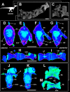
The origin of the air sac system present in birds has been an enigma for decades. Skeletal pneumaticity related to an air sac system is present in both derived non-avian dinosaurs and pterosaurs. But the question remained open whether this was a shared trait present in the common avemetatarsalian ancestor. We analyzed three taxa from the Late Triassic of South Brazil, which are some of the oldest representatives of this clade (233.23 ± 0.73 Ma), including two sauropodomorphs and one herrerasaurid. All three taxa present shallow lateral fossae in the centra of their presacral vertebrae. Foramina are present in many of the fossae but at diminutive sizes consistent with neurovascular rather than pneumatic origin. Micro-tomography reveals a chaotic architecture of dense apneumatic bone tissue in all three taxa. The early sauropodomorphs showed more complex vascularity, which possibly served as the framework for the future camerate and camellate pneumatic structures of more derived saurischians. Finally, the evidence of the absence of postcranial skeletal pneumaticity in the oldest dinosaurs contradicts the homology hypothesis for an invasive diverticula system and suggests that this trait evolved independently at least 3 times in pterosaurs, theropods, and sauropodomorphs.
© 2022. The Author(s).
Conflict of interest statement
The authors declare no competing interests.
Figures


Similar articles
-
Postcranial pneumaticity: an evaluation of soft-tissue influences on the postcranial skeleton and the reconstruction of pulmonary anatomy in archosaurs.J Morphol. 2006 Oct;267(10):1199-226. doi: 10.1002/jmor.10470. J Morphol. 2006. PMID: 16850471
-
Reassessment of the evidence for postcranial skeletal pneumaticity in Triassic archosaurs, and the early evolution of the avian respiratory system.PLoS One. 2012;7(3):e34094. doi: 10.1371/journal.pone.0034094. Epub 2012 Mar 28. PLoS One. 2012. PMID: 22470520 Free PMC article.
-
Osteological and Soft-Tissue Evidence for Pneumatization in the Cervical Column of the Ostrich (Struthio camelus) and Observations on the Vertebral Columns of Non-Volant, Semi-Volant and Semi-Aquatic Birds.PLoS One. 2015 Dec 9;10(12):e0143834. doi: 10.1371/journal.pone.0143834. eCollection 2015. PLoS One. 2015. PMID: 26649745 Free PMC article.
-
Air-filled postcranial bones in theropod dinosaurs: physiological implications and the 'reptile'-bird transition.Biol Rev Camb Philos Soc. 2012 Feb;87(1):168-93. doi: 10.1111/j.1469-185X.2011.00190.x. Epub 2011 Jul 7. Biol Rev Camb Philos Soc. 2012. PMID: 21733078 Review.
-
When the lung invades: a review of avian postcranial skeletal pneumaticity.Philos Trans R Soc Lond B Biol Sci. 2025 Feb 27;380(1920):rstb20230427. doi: 10.1098/rstb.2023.0427. Epub 2025 Feb 27. Philos Trans R Soc Lond B Biol Sci. 2025. PMID: 40010393 Free PMC article. Review.
Cited by
-
Macroevolutionary patterns in the pelvis, stylopodium and zeugopodium of megalosauroid theropod dinosaurs and their importance for locomotor function.R Soc Open Sci. 2023 Aug 16;10(8):230481. doi: 10.1098/rsos.230481. eCollection 2023 Aug. R Soc Open Sci. 2023. PMID: 37593714 Free PMC article.
-
Whence the birds: 200 years of dinosaurs, avian antecedents.Biol Lett. 2025 Jan;21(1):20240500. doi: 10.1098/rsbl.2024.0500. Epub 2025 Jan 22. Biol Lett. 2025. PMID: 39837495 Free PMC article. Review.
-
Variation in air sac morphology and postcranial skeletal pneumatization patterns in the African grey parrot.J Anat. 2025 Jan;246(1):1-19. doi: 10.1111/joa.14146. Epub 2024 Oct 7. J Anat. 2025. PMID: 39374322 Free PMC article.
-
The living dinosaur: accomplishments and challenges of reconstructing dinosaur physiology.Biol Lett. 2025 May;21(5):20250126. doi: 10.1098/rsbl.2025.0126. Epub 2025 May 29. Biol Lett. 2025. PMID: 40438994 Free PMC article. Review.
-
The influence of soft tissue volume on estimates of skeletal pneumaticity: implications for fossil archosaurs.Philos Trans R Soc Lond B Biol Sci. 2025 Feb 27;380(1920):20230428. doi: 10.1098/rstb.2023.0428. Epub 2025 Feb 27. Philos Trans R Soc Lond B Biol Sci. 2025. PMID: 40010389 Free PMC article.
References
-
- Proctor NS, Lynch PJ. Manual of Ornithology: Avian Structure & Function. Yale University Press; 1993.
-
- Britt BB, Makovicky PJ, Gauthier J, Bonde N. Postcranial pneumatization in Archaeopteryx. Nature. 1998;395:374–376. doi: 10.1038/26469. - DOI
MeSH terms
Grants and funding
- (FAPESP 2019/16727-3) 'Taphonomical landscapes'/Fundação de Amparo à Pesquisa do Estado de São Paulo
- (FAPESP 2019/16727-3) 'Taphonomical landscapes'/Fundação de Amparo à Pesquisa do Estado de São Paulo
- CNPq 307333/2021-3/Conselho Nacional de Desenvolvimento Científico e Tecnológico
- CNPq 404095/2021-6/Conselho Nacional de Desenvolvimento Científico e Tecnológico
- 422568/2018-0/Conselho Nacional de Desenvolvimento Científico e Tecnológico
- CNPq 307333/2021-3/Conselho Nacional de Desenvolvimento Científico e Tecnológico
- 21/2551-0000680-3/Fundação de Amparo à Pesquisa do Estado do Rio Grande do Sul
- FAPERGS 1/2551-0002030-0/Fundação de Amparo à Pesquisa do Estado do Rio Grande do Sul
- 21/2551-0000619-6/Fundação de Amparo à Pesquisa do Estado do Rio Grande do Sul
LinkOut - more resources
Full Text Sources
Miscellaneous

