

doi: 10.1038/s12276-022-00891-0.
Epub 2022 Dec 9.
SCON-a Short Conditional intrON for conditional knockout with one-step zygote injection
Affiliations
- PMID: 36494589
- PMCID: PMC9794761
- DOI: 10.1038/s12276-022-00891-0
Item in Clipboard
SCON-a Short Conditional intrON for conditional knockout with one-step zygote injection
Exp Mol Med.
2022 Dec.
Erratum in
-
Author Correction: SCON-a Short Conditional intrON for conditional knockout with one-step zygote injection.Exp Mol Med. 2023 Jun;55(6):1278-1280. doi: 10.1038/s12276-023-01039-4. Exp Mol Med. 2023. PMID: 37340143 Free PMC article. No abstract available.
Abstract
The generation of conditional alleles using CRISPR technology is still challenging. Here, we introduce a Short Conditional intrON (SCON, 189 bp) that enables the rapid generation of conditional alleles via one-step zygote injection. In this study, a total of 13 SCON mouse lines were successfully generated by 2 different laboratories. SCON has conditional intronic functions in various vertebrate species, and its target insertion is as simple as CRISPR/Cas9-mediated gene tagging.
© 2022. The Author(s).
Conflict of interest statement
The SCON technology described here is the subject of a patent application in which S.W. and B.K.K. are inventors.
Figures
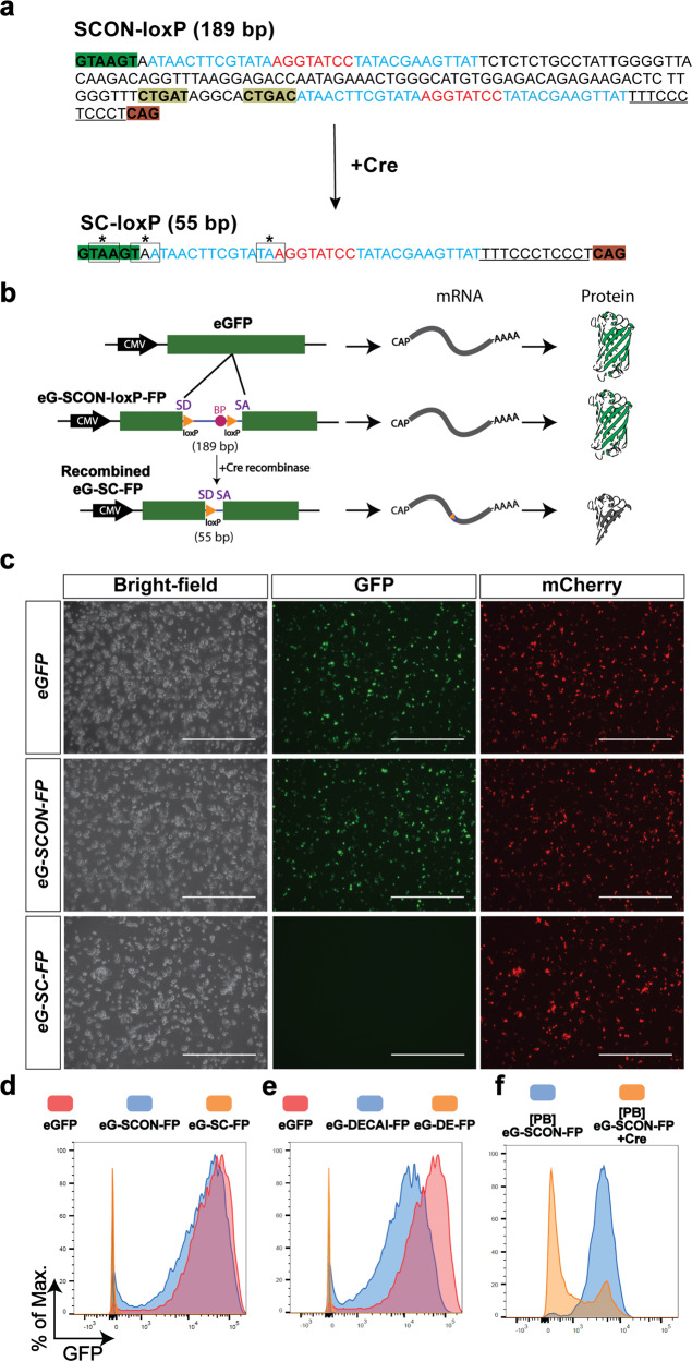
a Sequence of SCON before and after recombination. The intact SCON is 189 bp long, and the sequence is annotated as follows: nucleotides highlighted in green and dark orange at the two ends represent the splice donor and splice acceptor, respectively; nucleotides highlighted in dark yellow represent the putative branch points; sequences that are underlined represent the polypyrimidine tract; nucleotides in blue and red represent loxP sites consisting of 13 bp recognition sites and 8 bp spacers, respectively. In the recombined SCON form, which is 55 bp long, the remaining components include the splice donor, one loxP, polypyrimidine tract, and the splice acceptor; the three coding frames contain stop codons that are indicated by a box and an asterisk over the nucleotides. b Schematic diagram of the SCON functionality test in an eGFP overexpression construct including intact eGFP, eG-SCON-FP, and recombinant eG-SC-FP. SD, splice donor; BP, branch point; SA, splice acceptor. c Images of transfected HEK293T cells on Day 1 with intact eGFP, eG-SCON-FP and recombinant eG-SC-FP. All constructs were cotransfected with an mCherry overexpression plasmid. Scale bar, 1 mm. d, e Histograms of the flow cytometry analysis of transfected HEK293T cells showing comparisons between eGFP (red) and eG-SCON-FP (blue) (d), between eGFP (red) and eG-DECAI-FP (blue) (e), and the respective recombined forms eG-SC-FP and eG-DE-FP (yellow) (d, e). f Flow cytometry analysis of mouse ES cells with integrated piggyBac-eG-SCON-FP transfected with Cre-expressing plasmid (yellow) or empty vector (blue).
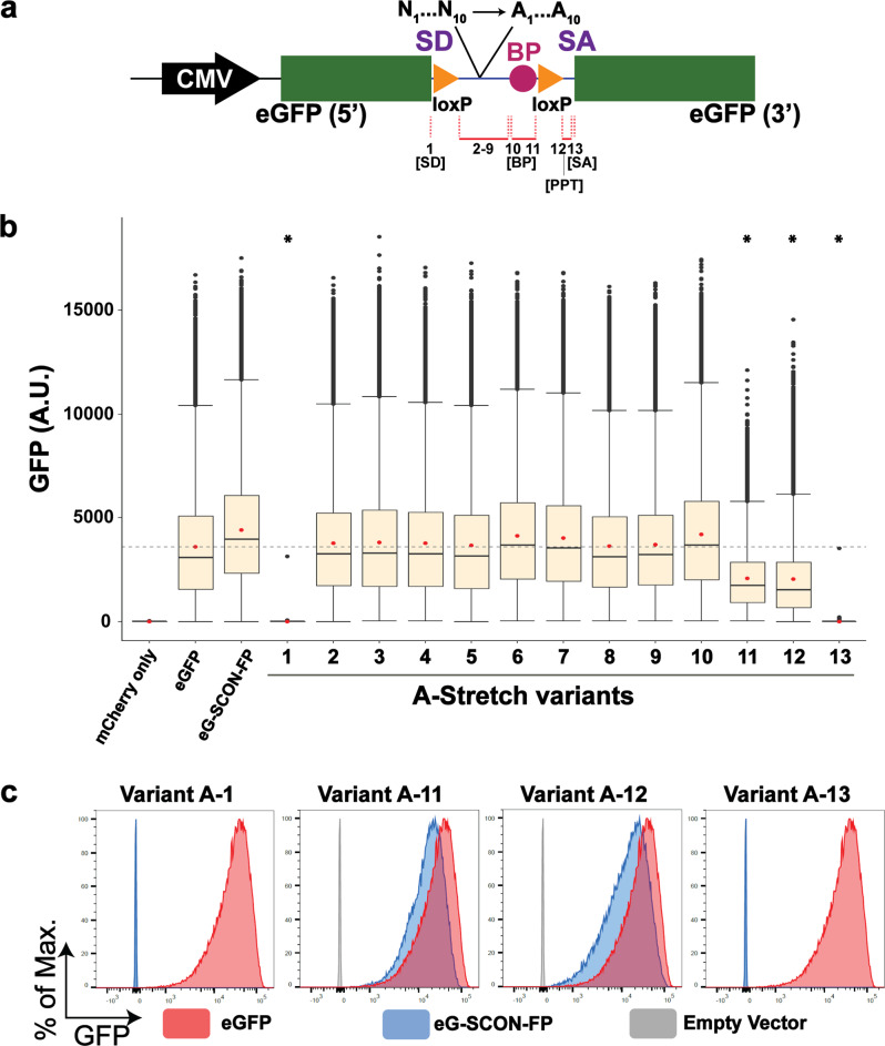
a Schematic diagram of the 13 A-stretch variants, where 6–10 nucleotides are converted to adenine, covering all sequences from SD to SA, excluding the LoxP sites. PPT, polypyrimidine tract. b Boxplots of measured scaled values from the flow cytometry analysis of HEK293T cells transfected with mCherry alone, eGFP, eG-SCON-FP, and the 13 A-stretch variants. The red dot indicates the mean. The gray horizontal dotted line indicates the mean value of eGFP. *indicates statistical significance (p = 0) from the unpaired t test with negative mean difference values when compared with the mean intensity value of eGFP. c Flow cytometry analysis of HEK293T cells transfected with variant A-1, A-11, A-12, or A-13 (blue) compared with intact eGFP (red) and the empty vector or mCherry alone (gray).
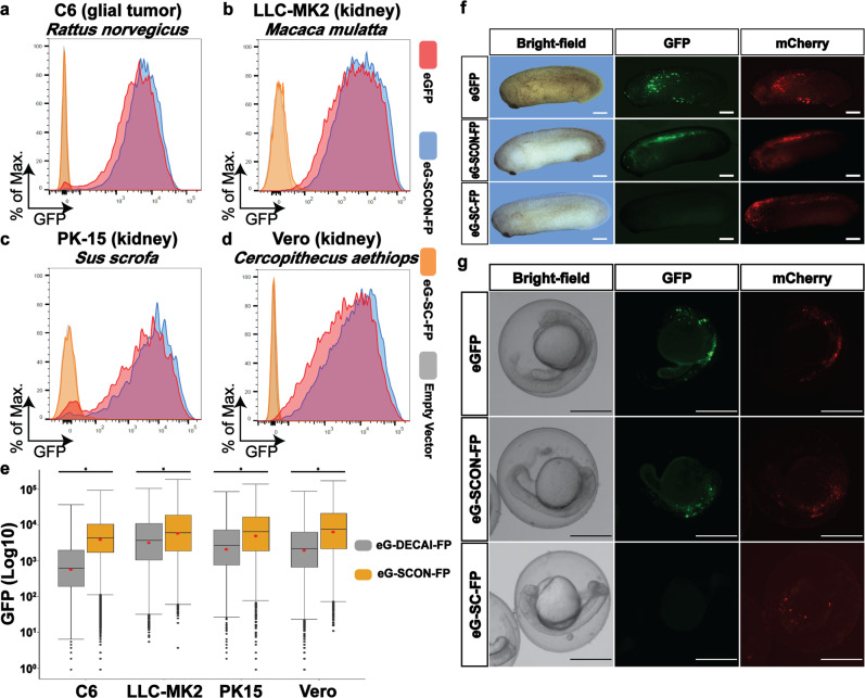
a–d Flow cytometry analysis of C6 (a), LLC-MK2 (b), PK-15 (c), and Vero (d) cells transfected with eGFP (red), eG-SCON-FP (blue), eG-SC-FP (yellow) or the empty vector or mCherry alone (gray). e Flow cytometry analysis comparing the GFP levels in various cell lines transfected with eG-DECAI-FP (gray) or eG-SCON-FP (yellow). *p < 0.001, from unpaired t test. f, g
Xenopus and zebrafish embryos injected with eGFP, eG-SCON-FP, or eG-SC-FP constructs, which were imaged 24 h postinjection. A plasmid containing mCherry was used as an injection control. Scale bar, 500 μm.
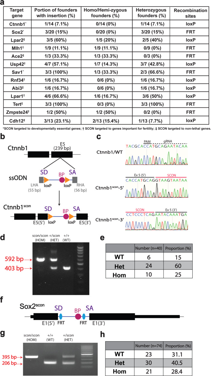
a List of alleles successfully targeted with SCON and the corresponding efficiencies, genotypes, and recombination sites used. b Schematic illustration of SCON targeting ssODN, with 55 and 56 bp left and right homology arms, respectively, in exon 5 of the Ctnnb1 gene. c Sanger sequencing track of the WT, 5′ and 3′ alleles of Ctnnb1 and Ctnnb1scon, respectively. d Genotyping PCR of Ctnnb1scon/scon (HOM), Ctnnb1+/scon (HET), and Ctnnb1+/+ (WT), in which the lower (403 bp) and upper (592 bp) bands correspond to the WT and knock-in alleles, respectively. e Genotype quantification from crosses of double heterozygotes (Ctnnb1+/scon). Total number of offspring, n = 40. f Schematic illustration of the Sox2scon allele. g Genotyping PCR of Sox2scon/scon (HOM), Sox2+/+ (WT), and Sox2+/scon (HET), in which the lower (206 bp) and upper (395 bp) bands correspond to the WT and knock-in alleles, respectively. h Genotyping quantification from crosses of heterozygotes (Sox2+/scon). Total number of offspring, n = 74.
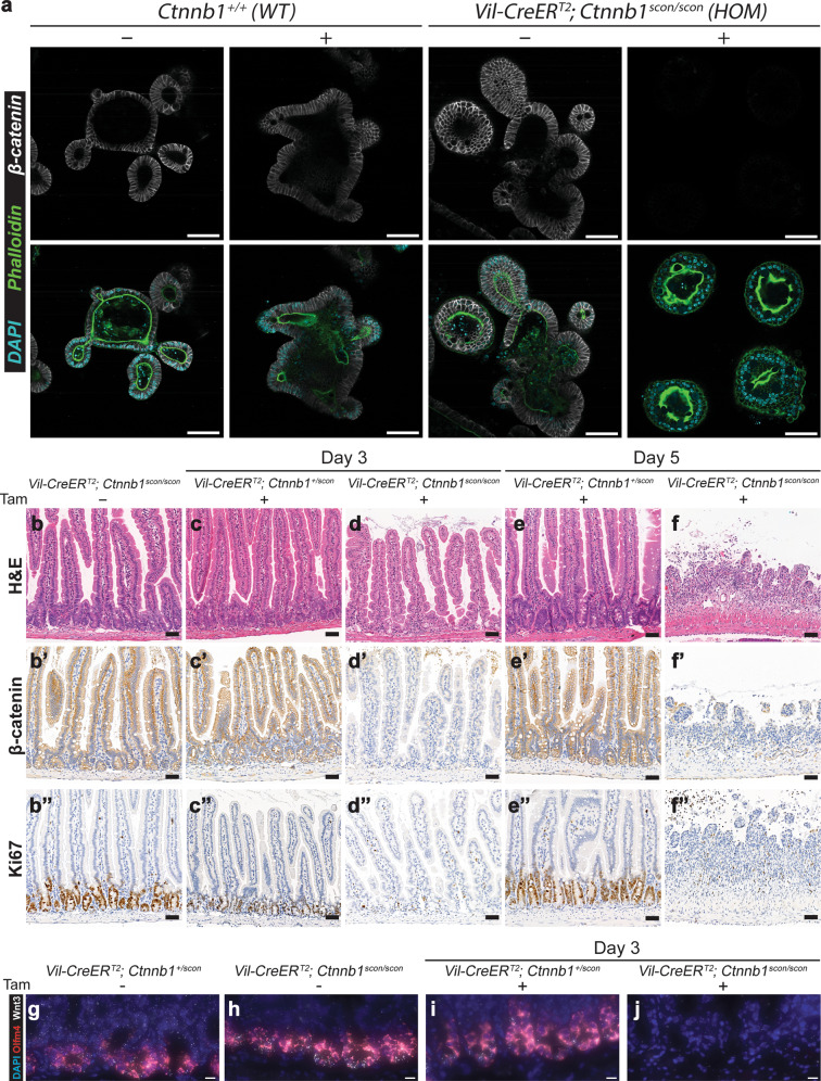
a Small intestinal organoids from WT (Ctnnb1+/+) and HOM (Vil-CreERT2; Ctnnb1scon/scon) mice treated with either 4-OH-tamoxifen (4-OHT) or vehicle for 8 h. Organoids were fixed on Day 4 and stained for β-catenin (gray), phalloidin (green) and DAPI (cyan). Scale bar, 100 μm. Homozygous (Vil-CreERT2; Ctnnb1scon/scon) intestines are healthy, with a normal epithelial crypt-villus morphology (b), β-catenin on the cell membrane (b′), and Ki67 marking proliferating cells in the crypts (b″). Heterozygous (Vil-CreERT2; Ctnnb1+/scon) intestines show no changes in morphology (c, e), β-catenin (c′, e) or Ki67 (c″, e″) after tamoxifen treatment. In homozygous intestines, tamoxifen treatment leads to the loss of crypts (d), β-catenin staining (d′), and Ki67 staining (d″) on Day 3. On Day 5, the epithelium is completely lost (f–f″). H&E, hematoxylin and eosin. Scale bar, 50 μm. g–j smRNA-FISH of intestinal sections with DAPI (blue), Olfm4 (red), and Wnt3 (white). g, h uninduced intestinal sections of HET (g) and HOM (h) mice; i, j Day 3 after induction with 3 mg tamoxifen per 20 g body weight of HET (i) and HOM (j) mice. Scale bar, 10 μm.
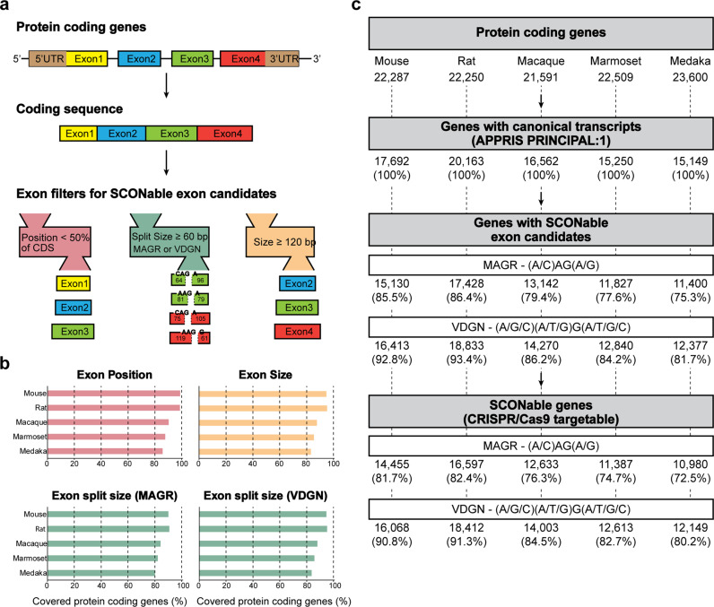
a Selection of the target exon candidates from protein-coding transcripts. b Gene coverage of individual exon filters, such as position, size, and split exon size. c Summary of databases of SCON insertion sites with CRISPR/Cas9 target sites.
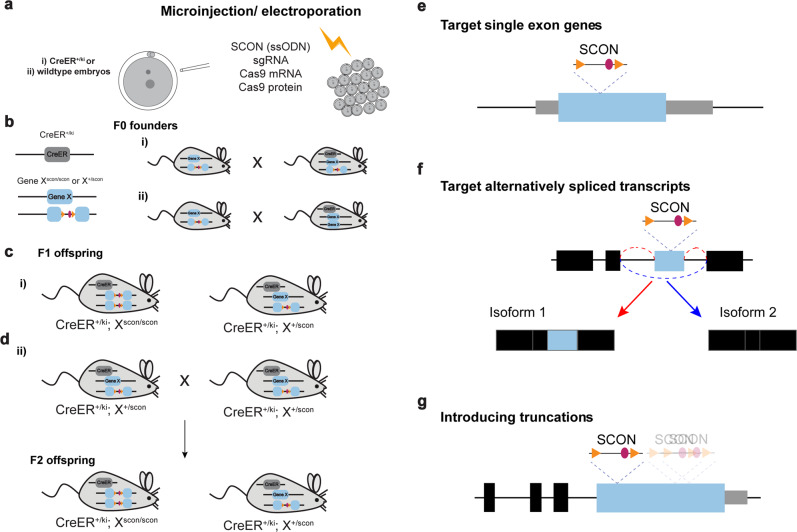
a SCON mouse generation in 1-cell-stage embryos via microinjection or electroporation. The embryos either (i) carry inducible CreER alleles or (ii) are wild-type. b The F0 founder mice are screened for SCON insertion in the target gene (gene X). Mice carrying a SCON insertion are crossed with each other (i) or are crossed with a CreER-bearing mouse (ii). c The resulting F1 offspring may contain genotypes ready for in vivo cKO studies (i). d The F1 offspring that carry a gene X-SCON allele as well as the CreER allele are crossed with each other to obtain experiment-ready cKO mice in the F2 generation (ii). e Illustration of the insertion of SCON in single-exon genes. f Illustration of the insertion of SCON into genes that express alternatively spliced transcripts. SCON can be inserted in the alternatively spliced exon, which would lead to the protein truncations produced by isoform 1, while isoform 2 is intact. g Illustration of the insertion of SCON in large exons (>1000 bp), which leads to various sizes of truncations.
References
Publication types
MeSH terms
LinkOut - more resources
Full Text Sources
Research Materials
