

The incidence of radicular groove on maxillary lateral incisors of Saudi population: CBCT evaluation
- PMID: 36494650
- PMCID: PMC9733403
- DOI: 10.1186/s12903-022-02616-1
The incidence of radicular groove on maxillary lateral incisors of Saudi population: CBCT evaluation
Abstract
Background: The radicular groove (RG) is one of the developmental anomalies that is commonly found in maxillary incisors. The formation of radicular groove is initiated around the cingulum and can reach the root at different levels. The incidence of radicular grooves was reported in different countries but there was no published data about the incidence of RG in Saudi Arabia. Therefore, this study aimed to evaluate the incidence of radicular grooves on maxillary lateral incisors in the Saudi population using cone-beam computed tomography (CBCT).
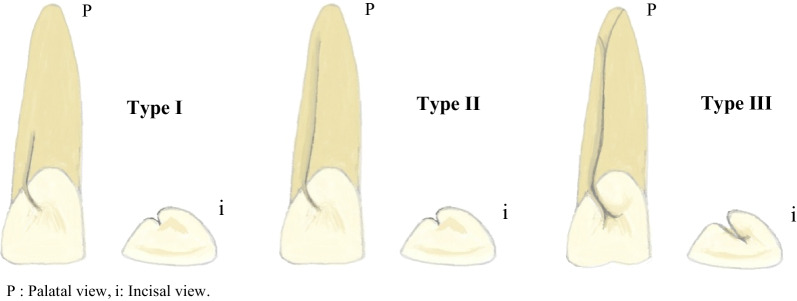
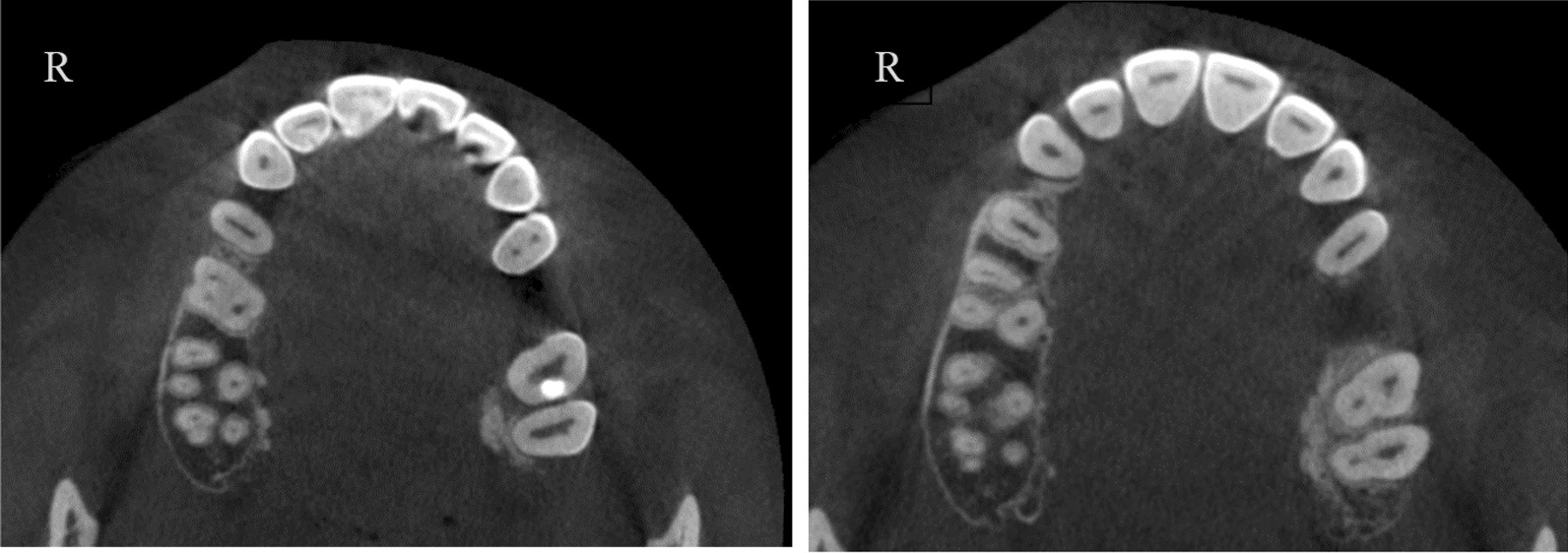
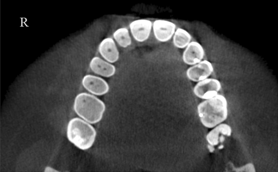
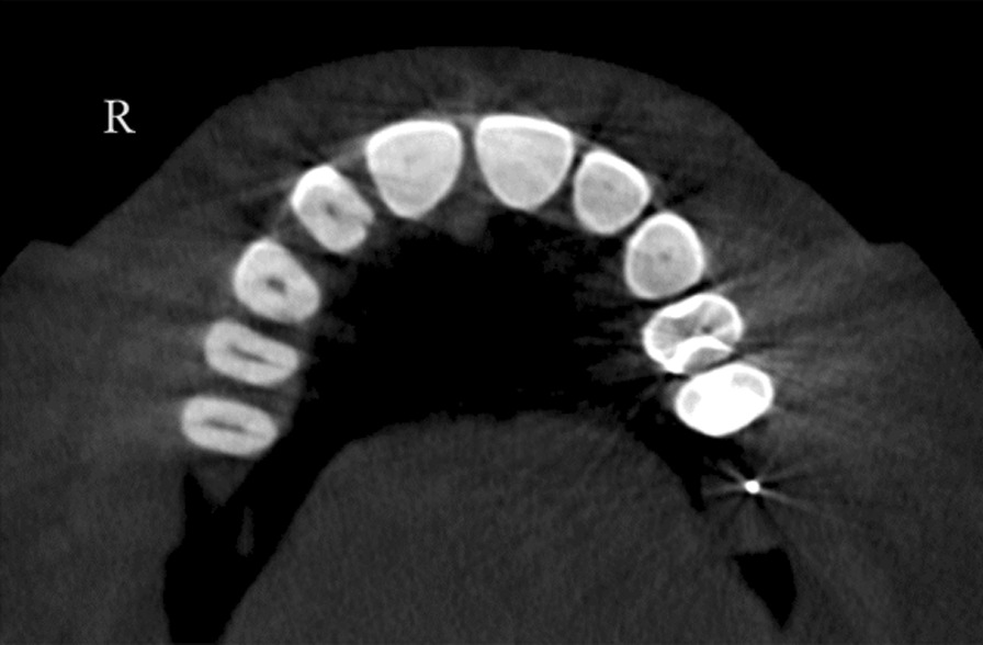
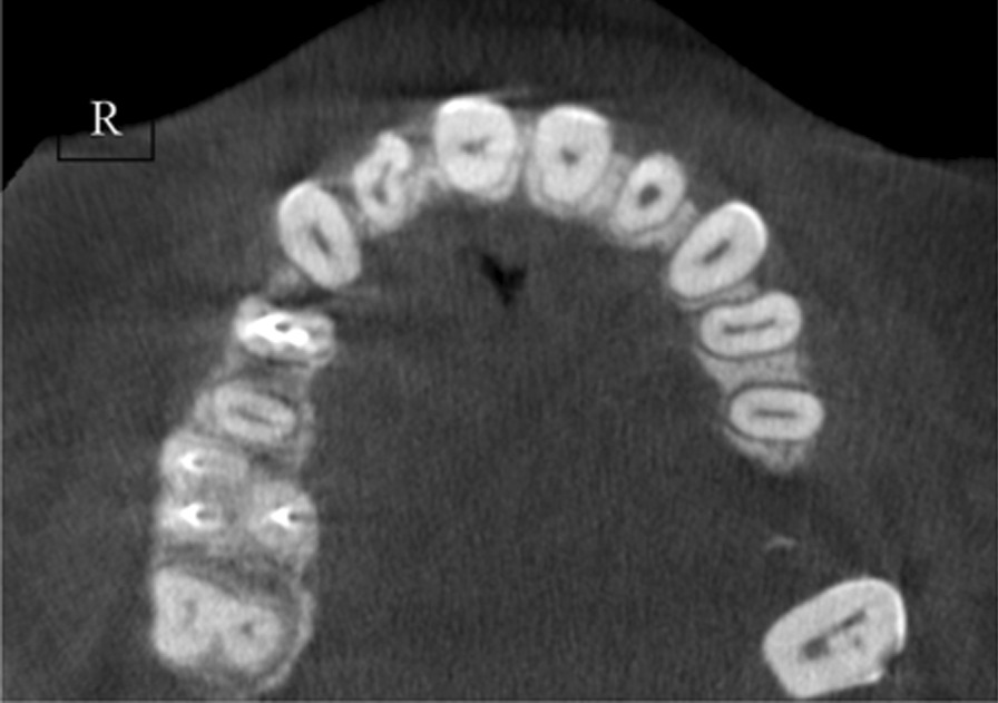
Methods: The dental records of 490 patients (N = 490) with CBCT images of maxillary anterior teeth were screened for inclusion criteria. Then 264 included cases were evaluated independently by two Endodontists. The evaluation was performed on CBCT images in the axial, sagittal, and coronal sections using Planmeca Romexis® software. The following data were recorded for each patient: Patients' age and gender, radicular groove presence or absence, and if it is bilateral or unilateral. The type of radicular groove was recorded according to Gu's classification (type I, II, or III).
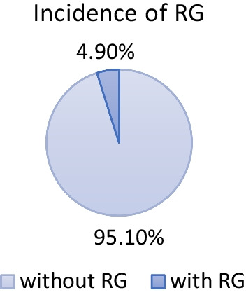
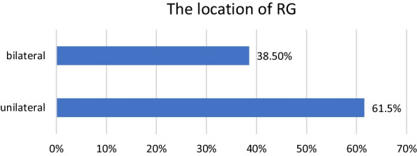
Results: The incidence rate of radicular grooves in maxillary lateral incisors was 4.9%. RG was found to be unilateral in 61.5% and bilateral in 38.5%. The majority of RG were classified as type I in 69.2%, followed by type II in 15.4%, and type III was found in 15.4%.
Conclusion: 4.9% of the Saudi population has RG in the upper lateral incisor. This anatomical variation is mostly present as type I on one side only (unilateral).
Keywords: Dental anomaly; Endodontics; Palatal groove; Radicular groove.
© 2022. The Author(s).
Conflict of interest statement
The authors have no relevant financial or non-financial interests to disclose.
Figures
References
-
- Black GV. Operative dentistry: pathology of the hard tissues of teeth. 7. Chicago: Medico-Dental Publishing Company; 1936.
-
- Lee KW, Lee EC, Poon KY. Palato-gingival grooves in maxillary incisors. A possible predisposing factor to localized periodontal disease. Br Dent J. 1968;124:14–18. - PubMed
MeSH terms
LinkOut - more resources
Full Text Sources
