. 2022 Dec 9;17(1):431.
doi: 10.1186/s13023-022-02582-5.

Clinical and genetic characteristics of Chinese patients with congenital cranial dysinnervation disorders

Affiliations

Clinical and genetic characteristics of Chinese patients with congenital cranial dysinnervation disorders

Hongyan Jia et al. Orphanet J Rare Dis. .

Abstract

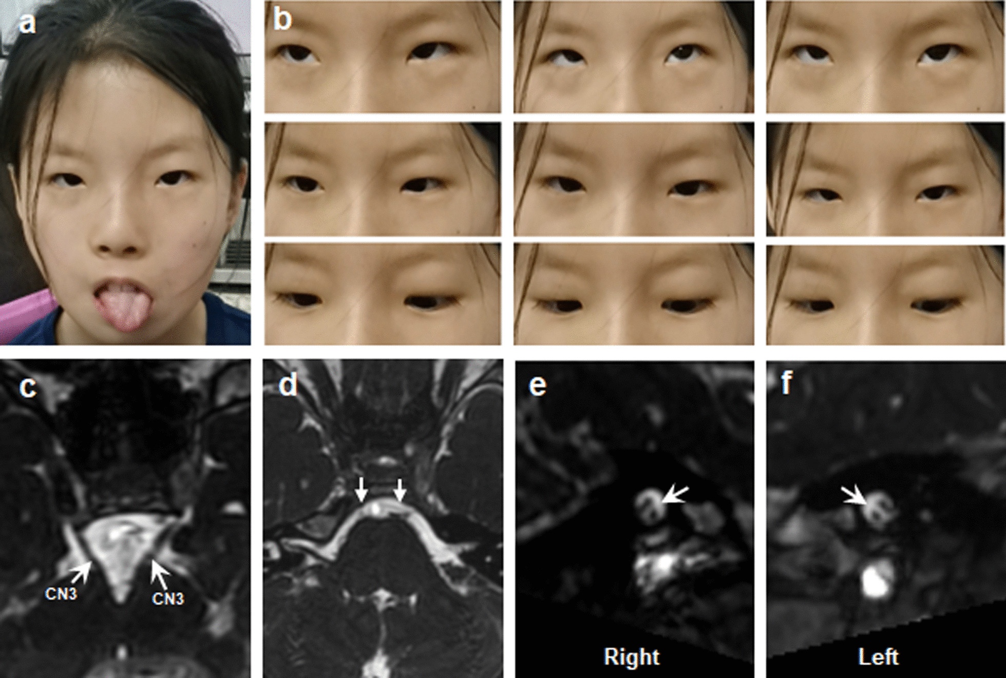
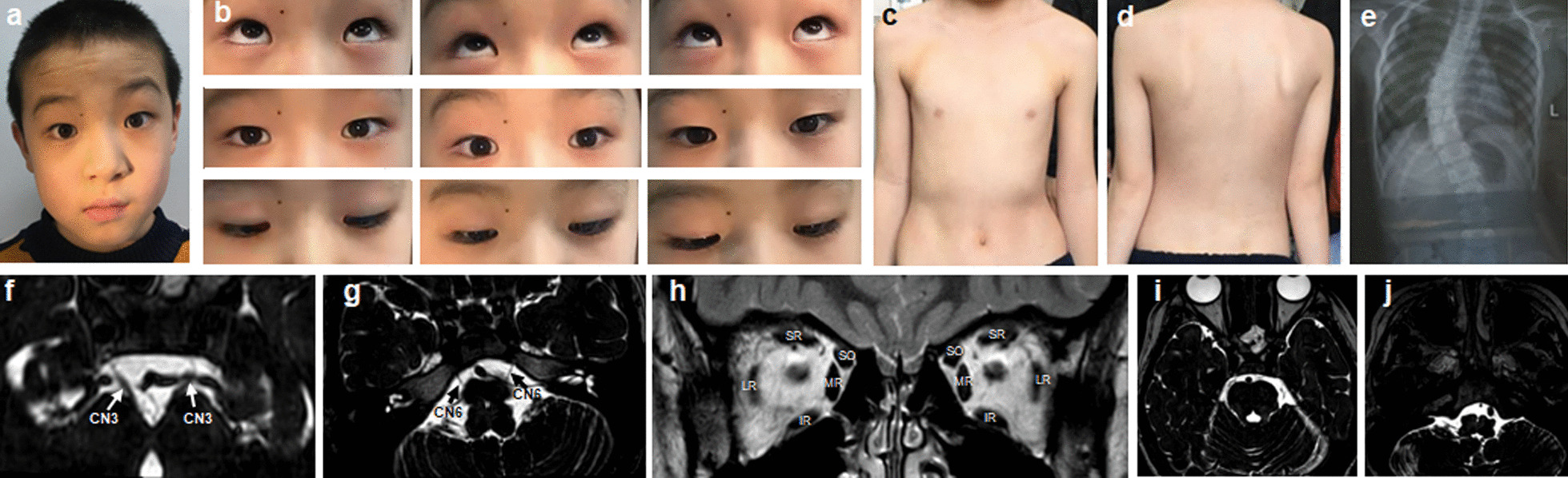
Background: Congenital cranial dysinnervation disorders (CCDDs) are a group of diseases with high clinical and genetic heterogeneity. Clinical examinations combined with Magnetic resonance imaging (MRI) and whole exome sequencing (WES) were performed to reveal the phenotypic and genotypic characteristics in a cohort of Chinese CCDDs patients.

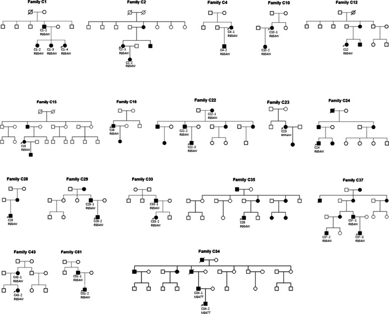
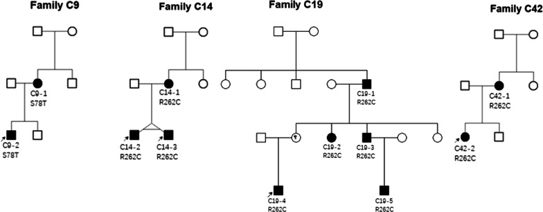
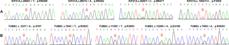
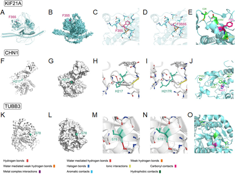
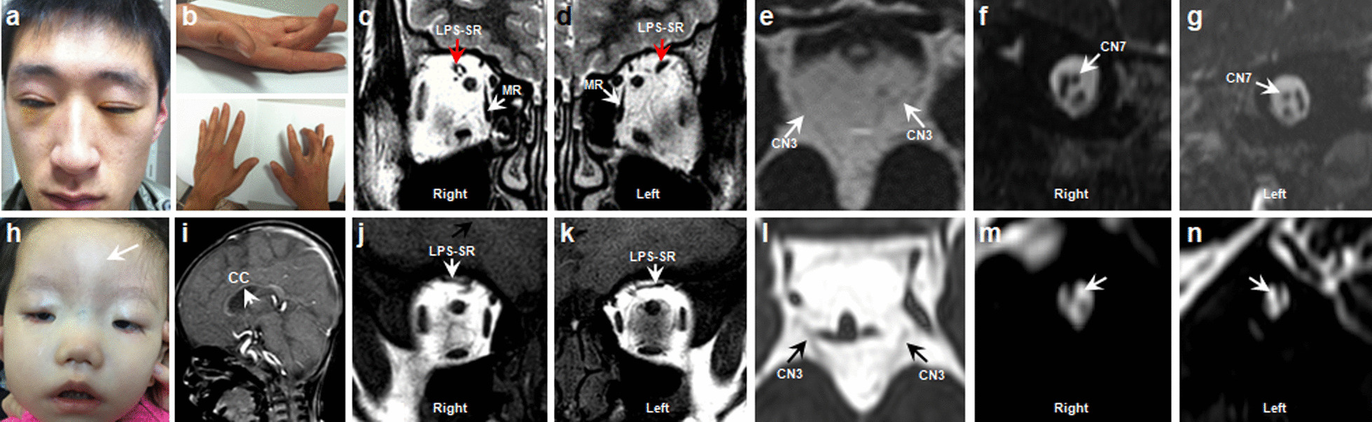
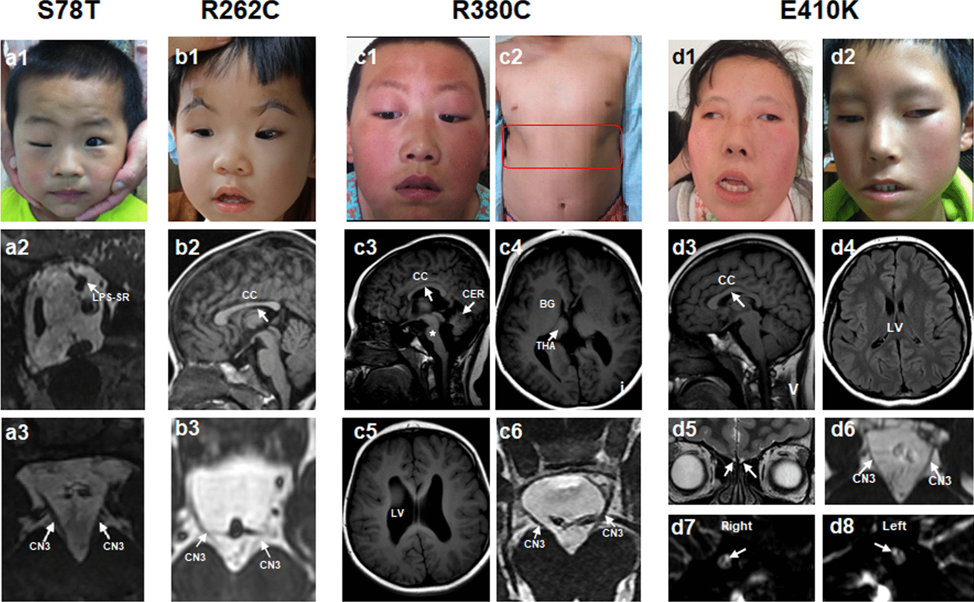
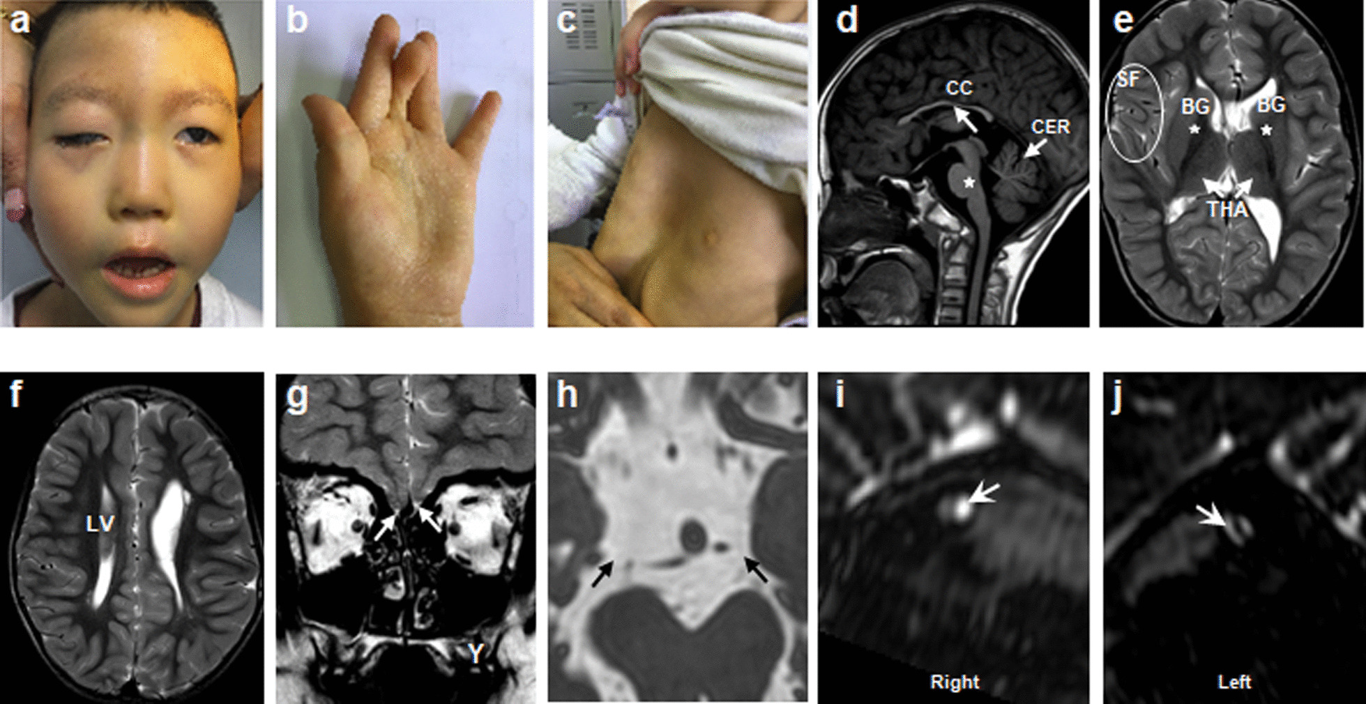
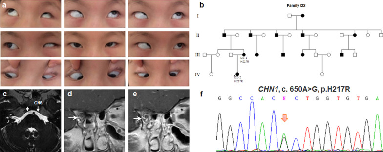
Results: A total of 122 CCDDs patients from 96 families were enrolled. All patients showed restrictive eye movements, and 46 patients from 46 families (47.9%, 46/96) were accompanied by multiple congenital malformations. Multi-positional high-resolution MRI was performed in 94 patients from 88 families, of which, all patients had hypoplasia of the cranial nerves except HGPPS patients and 15 patients from 15 families (17.0%,15/88) were accompanied by other craniocerebral malformations. WES was performed in 122 CCDDs patients. Ten pathogenic variants were detected in KIF21A, TUBB3, and CHN1 genes in 43 families. Three variants were unreported, including KIF21A (c.1064T > C, p.F355S), TUBB3 (c.232T > A, p.S78T) and CHN1 (c.650A > G, p.H217R). Of the 43 probands harboring pathogenic variants, 42 were diagnosed with Congenital Fibrosis of Extraocular Muscles (CFEOM) and one was Duane Retraction Syndrome (DRS). No definite pathogenic variants in known candidate genes of CCDDs were found in sporadic DRS, Möbius Syndrome (MBS) and Horizontal Gaze Palsy with Progressive Scoliosis (HGPPS) patients. The CFEOM patients harboring R380C, E410K and R262H variants in TUBB3 gene and F355S variant in KIF21A gene exhibited syndromic phenotypes.

Conclusions: This study broadened the phenotypic and genotypic spectrums of CCDDs, and it was the largest clinical and genetic investigation for CCDDs patients from China. KIF21A and TUBB3 were the common pathogenic genes in Chinese CFEOM. MRI coupled with WES can provide a supportive diagnosis in patients with clinically suspected CCDDs.

Keywords: Congenital cranial dysinnervation disorders; Genetics; Magnetic resonance imaging; Phenotypic and genotypic characteristics; Whole exome sequencing.


Conflict of interest statement

The authors declare that they have no competing interests.

Figures

Fig. 1
Fig. 1
The family trees of familial forms CFEOM patients carrying KIF21A variants
Fig. 2
Fig. 2
The family trees of familial forms CFEOM patients carrying TUBB3 variants
Fig. 3
Fig. 3
Genomic sequence chromatograms of CFEOM individuals with KIF21A and TUBB3 variants. A Four heterozygous variants (R954W, R954Q, M947T and F355S) of KIF21A gene (red arrows). B Five heterozygous variants (S78T, R262C, R380C, E410K and R262H) of TUBB3 gene (red arrows)
Fig. 4
Fig. 4
Spatial distribution of protein key sites and changes of amino acid interaction force before and after mutation. A, B KIF21A protein structure (cartoon/surface form) and spatial distribution of F355 residue. C, D Interaction force analysis of the KIF21A F355 and KIF21A F355S with the surrounding amino acid residues. E Showing positions of F355 and previously reported variants associated with CFEOM in the motor domain. F, G CHN1 protein structure (cartoon/surface form) and spatial distribution of H217 residue. H, I Interaction force analysis of the CHN1 H217 and CHN1 H217R with the surrounding amino acid residues. J Showing positions of H217 and other nearby previously reported variants in the same domain. K, L TUBB3 protein structure (cartoon/surface form) and spatial distribution of S78 residue. M, N Interaction force analysis of the TUBB3 S78 and TUBB3 S78T with the surrounding amino acid residues. O Showing positions of S78 and other nearby previously reported variants in the same domain
Fig. 5
Fig. 5
Clinical spectrum and MRI findings of patient C3 (ag) and C46 (hn). a, b Photographs of patient C3 harboring KIF21A R954W shows congenital bilateral ptosis (after correction of blepharoptosis) and joint contracture of right middle finger. cg MRI findings of patient C3 (The MRI was taken at the age of 16). Atrophy of bilateral levator palpebrae superioris-superior rectus (LPS-SR) and bilateral medial rectus (MR), hypoplasia of bilateral oculomotor nerves (CN3), and bilateral facial nerves (CN7) are normal (f, g). h Facial photograph of patient C46 harboring KIF21A F355S shows congenital bilateral ptosis, facial weakness and bony prominence (arrow). in MRI findings of patient C3 (The MRI was taken at the age of 1y). Hypoplasia of corpus callosum (CC), LPS-SR, CN3 and absence of CN7(mn, arrow)
Fig. 6
Fig. 6
Clinical and MRI spectrum of CFEOM patients with TUBB3 variants. (a1a3), Patient C9-2 harboring S78T variant. a1 Congenital ptosis of right eye. a2 Atrophy of levator palpebrae superioris-superior rectus (arrow) in right eye. a3 Hypoplasia of the CN3 at the brainstem. The MRI was taken at the age of 4y. b1b3 Patient C65 harboring R262C variant. b1 Congenital bilateral ptosis. b2, b3 Hypoplasia of CC and CN3. The MRI was taken at the age of 1y. c1c6 Patient C6 harboring R380C variant. c1, c2 Congenital bilateral ptosis and chest wall malformation (red box). c3, c4 Hypoplasia of CC, brainstem (c, star), cerebellum (CER), basal ganglia (BG), thalamus (THA) and CN3 with the enlargement of lateral ventricle (LV). The MRI was taken at the age of 11y. d1d8 Patients harboring E410K variants. d1, d2 Facial pictures of two patients (C21 and C32). d3d8 Patient C32 exhibits hypoplasia of CC (d3), asymmetry of LV (d4), absence of olfactory bulbs and sulci (d5, arrow), hypoplasia of CN3 (d6) and absence of bilateral CN7 (d7, d8, arrow). The MRI was taken at the age of 19y
Fig. 7
Fig. 7
Clinical spectrum and MRI findings of CFEOM patients with TUBB3 R262H variant. ac Congenital bilateral ptosis, facial weakness, joint contracture and funnel chest. df Hypoplasia of CC, brainstem (d, star), CER, BG, THA with asymmetry of Sylvian fissure (SF) and LV. gj Absence of olfactory bulbs and sulci (g, arrow), CN3 (h, arrow) and CN7 (i, j, arrow). The MRI was taken at the age of 4y
Fig. 8
Fig. 8
Photographs of the patient D2-1 harboring H217R variant in CHN1 gene. a Nine gaze photographs show limited abduction, globe retraction with narrowing of the palpebral fissure on attempted adduction. b Pedigree of autosomal dominant D2. ce Hypoplasia of the bilateral CN6 and aberrant innervation of LR by the inferior branches of CN3. d, e Two consecutive coronal MRI images to show the course of CN3. The MRI was taken at the age of 5y. f Genomic sequence chromatogram of heterozygous variant c.650A > G, p.H217R (arrow) in CHN1 gene
Fig. 9
Fig. 9
Clinical spectrum and MRI findings of patient M11 with MBS. a Facial photograph shows bilateral congenital facial weakness and hypoplasia of tongue. b Nine gaze photographs show limited eye movements. cf MRI findings of patient C3 (The MRI was taken at the age of 9). Normal CN3 and hypoplasia of the bilateral CN6 and CN7 (cf, arrow)
Fig. 10
Fig. 10
Clinical spectrum, MRI findings and spine X-ray of patient H4 with HGPPS. a Facial photograph. b Nine gaze photographs show limited horizontal eye movements and full vertical eye movements. c, d Photographs of the body show obvious scoliosis. e Spine X-ray shows obvious thoracolumbar scoliosis. fh Normal CN3, CN6 and EOMs. The MRI was taken at the age of 9y. i A deep posterior midsagittal cleft of the pons. j Flattening and butterfly-like morphology in the medulla

References

    1. Gutowski NJ, Bosley TM, Engle EC. 110th ENMC International Workshop: the congenital cranial dysinnervation disorders (CCDDs). Naarden, The Netherlands, 25–27 October, 2002. Neuromuscul Disord 2003;13(7–8):573–8. - PubMed
    1. Chilton JK, Guthrie S. Axons get ahead: Insights into axon guidance and congenital cranial dysinnervation disorders. Dev Neurobiol. 2017;77(7):861–875. doi: 10.1002/dneu.22477. - DOI - PubMed
    1. MacKinnon S, Oystreck DT, Andrews C, Chan WM, Hunter DG, Engle EC. Diagnostic distinctions and genetic analysis of patients diagnosed with moebius syndrome. Ophthalmology. 2014;121(7):1461–1468. doi: 10.1016/j.ophtha.2014.01.006. - DOI - PMC - PubMed
    1. Whitman MC, Barry BJ, Robson CD, Facio FM, Van Ryzin C, Chan WM, et al. TUBB3 R262H causes a recognizable syndrome including CFEOM3, facial palsy, joint contractures, and early-onset peripheral neuropathy. Hum Genet. 2021;140(12):1709–1731. doi: 10.1007/s00439-021-02379-9. - DOI - PMC - PubMed
    1. Yamada K, Andrews C, Chan WM, McKeown CA, Magli A, et al. Heterozygous mutations of the kinesin KIF21A in congenital fibrosis of the extraocular muscles type 1 (CFEOM1) Nat Genet. 2003;35(4):318–321. doi: 10.1038/ng1261. - DOI - PubMed

Publication types

LinkOut - more resources