

Hox gene functions in the C. elegans nervous system: From early patterning to maintenance of neuronal identity
- PMID: 36496326
- PMCID: PMC10244487
- DOI: 10.1016/j.semcdb.2022.11.012
Hox gene functions in the C. elegans nervous system: From early patterning to maintenance of neuronal identity
Abstract
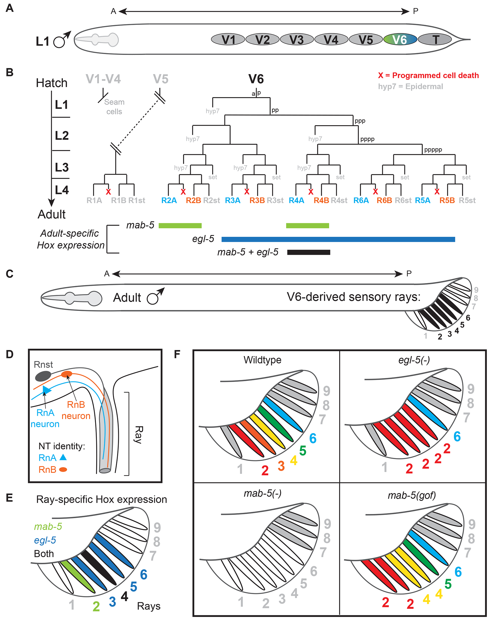
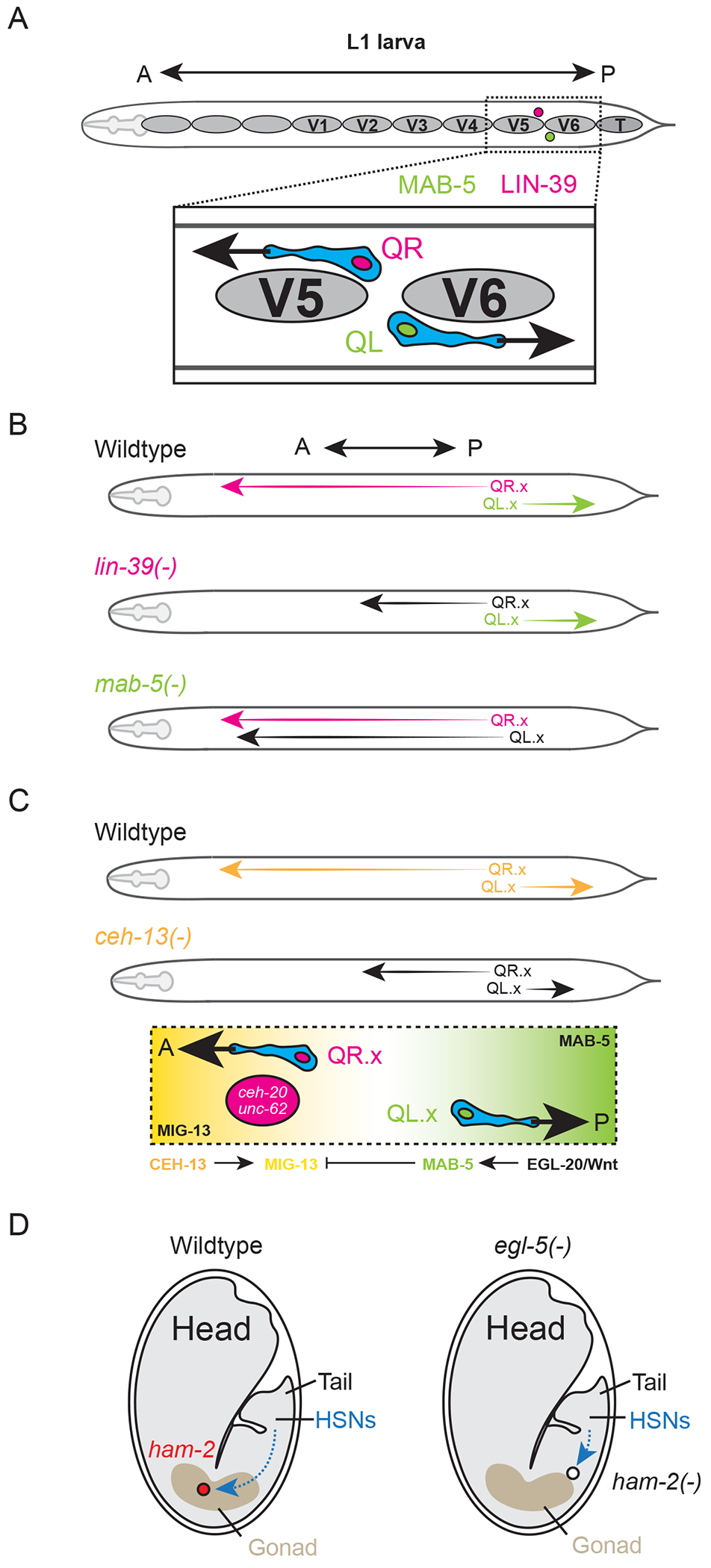
The nervous system emerges from a series of genetic programs that generate a remarkable array of neuronal cell types. Each cell type must acquire a distinct anatomical position, morphology, and function, enabling the generation of specialized circuits that drive animal behavior. How are these diverse cell types and circuits patterned along the anterior-posterior (A-P) axis of the animal body? Hox genes encode transcription factors that regulate cell fate and patterning events along the A-P axis of the nervous system. While most of our understanding of Hox-mediated control of neuronal development stems from studies in segmented animals like flies, mice, and zebrafish, important new themes are emerging from work in a non-segmented animal: the nematode Caenorhabditis elegans. Studies in C. elegans support the idea that Hox genes are needed continuously and across different life stages in the nervous system; they are not only required in dividing progenitor cells, but also in post-mitotic neurons during development and adult life. In C. elegans embryos and young larvae, Hox genes control progenitor cell specification, cell survival, and neuronal migration, consistent with their neural patterning roles in other animals. In late larvae and adults, C. elegans Hox genes control neuron type-specific identity features critical for neuronal function, thereby extending the Hox functional repertoire beyond early patterning. Here, we provide a comprehensive review of Hox studies in the C. elegans nervous system. To relate to readers outside the C. elegans community, we highlight conserved roles of Hox genes in patterning the nervous system of invertebrate and vertebrate animals. We end by calling attention to new functions in adult post-mitotic neurons for these paradigmatic regulators of cell fate.
Keywords: C. elegans; Cell migration; Cell survival; Hox transcription factors; Neural progenitors; Neuronal terminal identity.
Copyright © 2022 The Authors. Published by Elsevier Ltd.. All rights reserved.
Conflict of interest statement
Declaration of Competing Interest The authors declare no interests or relationships - financial, personal, or otherwise - that influence or could influence the work reported in this paper.
Figures


Similar articles
-
Emerging Roles for Hox Proteins in the Last Steps of Neuronal Development in Worms, Flies, and Mice.Front Neurosci. 2022 Feb 4;15:801791. doi: 10.3389/fnins.2021.801791. eCollection 2021. Front Neurosci. 2022. PMID: 35185450 Free PMC article. Review.
-
Nervous system-wide analysis of Hox regulation of terminal neuronal fate specification in Caenorhabditis elegans.PLoS Genet. 2022 Feb 28;18(2):e1010092. doi: 10.1371/journal.pgen.1010092. eCollection 2022 Feb. PLoS Genet. 2022. PMID: 35226663 Free PMC article.
-
Maintenance of neuronal identity in C. elegans and beyond: Lessons from transcription and chromatin factors.Semin Cell Dev Biol. 2024 Feb 15;154(Pt A):35-47. doi: 10.1016/j.semcdb.2023.07.001. Epub 2023 Jul 11. Semin Cell Dev Biol. 2024. PMID: 37438210 Free PMC article. Review.
-
Caenorhabditis elegans embryonic axial patterning requires two recently discovered posterior-group Hox genes.Proc Natl Acad Sci U S A. 2000 Apr 25;97(9):4499-503. doi: 10.1073/pnas.97.9.4499. Proc Natl Acad Sci U S A. 2000. PMID: 10781051 Free PMC article.
-
A map of terminal regulators of neuronal identity in Caenorhabditis elegans.Wiley Interdiscip Rev Dev Biol. 2016 Jul;5(4):474-98. doi: 10.1002/wdev.233. Epub 2016 May 2. Wiley Interdiscip Rev Dev Biol. 2016. PMID: 27136279 Free PMC article. Review.
Cited by
-
Chondrodystrophic Dogs as a Preclinical Large Animal Model of Discogenic Back Pain.JOR Spine. 2025 Jul 14;8(3):e70082. doi: 10.1002/jsp2.70082. eCollection 2025 Sep. JOR Spine. 2025. PMID: 40662113 Free PMC article.
-
Investigating the Functions of Hox Genes Using Planarian Asexual Reproduction.Methods Mol Biol. 2025;2889:91-106. doi: 10.1007/978-1-0716-4322-8_7. Methods Mol Biol. 2025. PMID: 39745607
-
VAB-8/KIF26, LIN-17/Frizzled, and EFN-4/Ephrin, control distinct stages of posterior neuroblast migration downstream of the MAB-5/Hox transcription factor in Caenorhabditis elegans.bioRxiv [Preprint]. 2025 May 29:2025.05.28.656589. doi: 10.1101/2025.05.28.656589. bioRxiv. 2025. PMID: 40501873 Free PMC article. Preprint.
-
Effects of HOXC8 on the Proliferation and Differentiation of Porcine Preadipocytes.Animals (Basel). 2023 Aug 14;13(16):2615. doi: 10.3390/ani13162615. Animals (Basel). 2023. PMID: 37627406 Free PMC article.
-
Pervasive homeobox gene function in the male-specific nervous system of Caenorhabditis elegans.bioRxiv [Preprint]. 2025 May 17:2025.05.13.653874. doi: 10.1101/2025.05.13.653874. bioRxiv. 2025. Update in: Development. 2025 Aug 15;152(16):dev204958. doi: 10.1242/dev.204958. PMID: 40463159 Free PMC article. Updated. Preprint.
References
-
- Hobert O, Kratsios P, Neuronal identity control by terminal selectors in worms, flies, and chordates, Current Opinion in Neurobiology 56 (2019) 97–105. - PubMed
-
- Hobert O, Chapter Twenty-Five - Terminal Selectors of Neuronal Identity, in: Wassarman PM (Ed.), Current Topics in Developmental Biology, Academic Press; 2016, pp. 455–475. - PubMed
-
- Di Bonito M, Glover JC, Studer M, Hox genes and region-specific sensorimotor circuit formation in the hindbrain and spinal cord, Developmental Dynamics 242(12) (2013) 1348–1368. - PubMed
Publication types
Grants and funding
LinkOut - more resources
Full Text Sources
