. 2022 Dec 10;13(1):7636.
doi: 10.1038/s41467-022-35169-6.

CRL2ZER1/ZYG11B recognizes small N-terminal residues for degradation

Affiliations

CRL2ZER1/ZYG11B recognizes small N-terminal residues for degradation

Yao Li et al. Nat Commun. .

Abstract

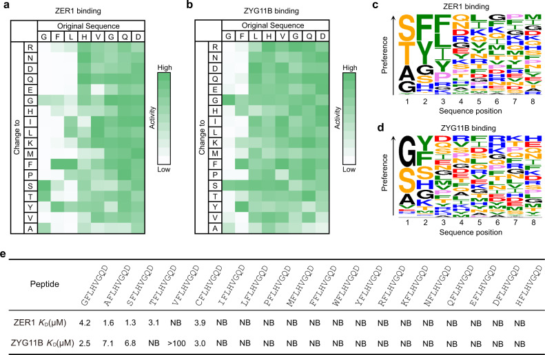
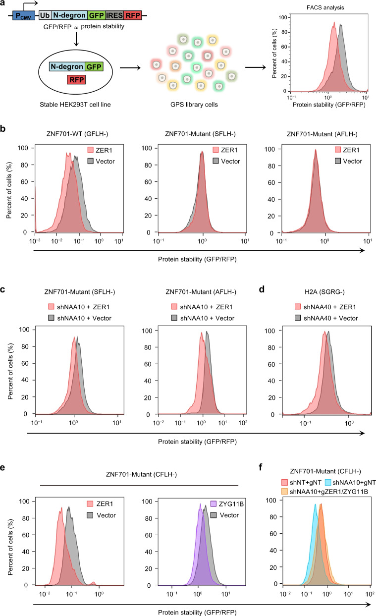
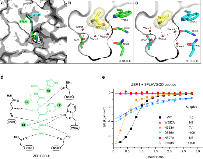
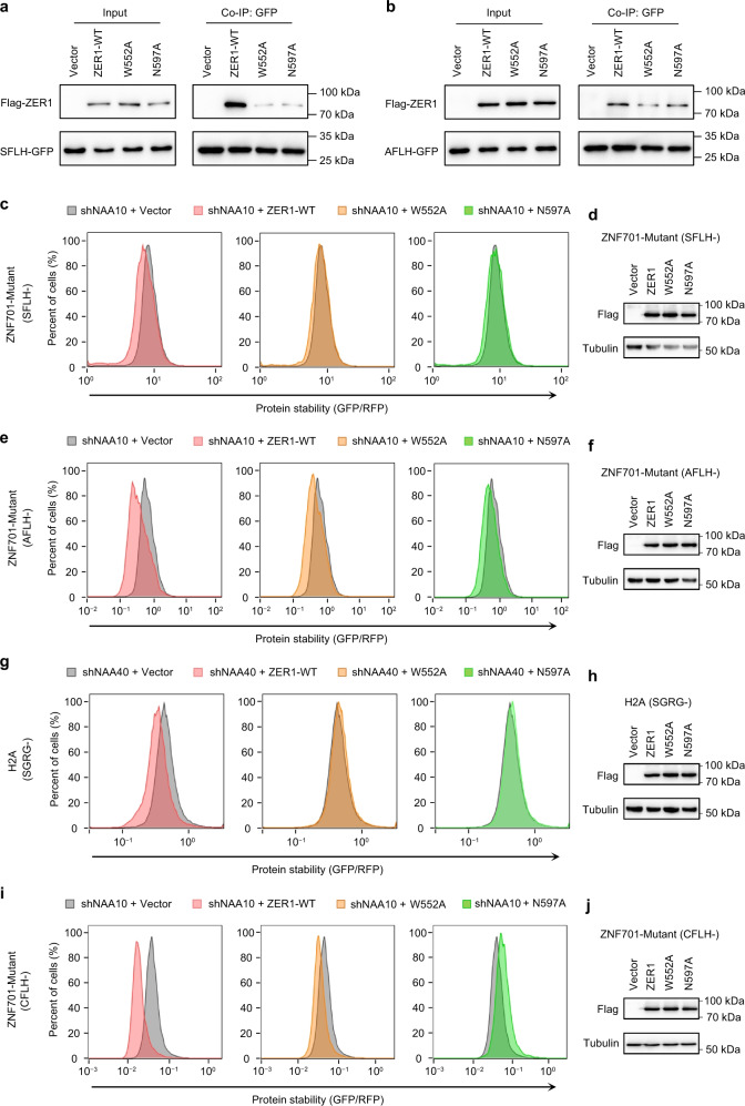
N-degron pathway plays an important role in the protein quality control and maintenance of cellular protein homeostasis. ZER1 and ZYG11B, the substrate receptors of the Cullin 2-RING E3 ubiquitin ligase (CRL2), recognize N-terminal (Nt) glycine degrons and participate in the Nt-myristoylation quality control through the Gly/N-degron pathway. Here we show that ZER1 and ZYG11B can also recognize small Nt-residues other than glycine. Specifically, ZER1 binds better to Nt-Ser, -Ala, -Thr and -Cys than to -Gly, while ZYG11B prefers Nt-Gly but also has the capacity to recognize Nt-Ser, -Ala and -Cys in vitro. We found that Nt-Ser, -Ala and -Cys undergo Nt-acetylation catalyzed by Nt-acetyltransferase (NAT), thereby shielding them from recognition by ZER1/ZYG11B in cells. Instead, ZER1/ZYG11B readily targets a selection of small Nt-residues lacking Nt-acetylation for degradation in NAT-deficient cells, implicating its role in the Nt-acetylation quality control. Furthermore, we present the crystal structures of ZER1 and ZYG11B bound to various small Nt-residues and uncover the molecular mechanism of non-acetylated substrate recognition by ZER1 and ZYG11B.


Conflict of interest statement

The authors declare no competing interests.

Figures

Fig. 1
Fig. 1. ZER1 and ZYG11B can recognize non-glycine small Nt-residues.
a, b Binding patterns of GST-tagged ZER1469–766 and ZYG11B490–744 to peptide SPOT arrays containing the GFLHVGQDGLELPTS peptide and its indicated derivatives containing mutations in the first 8 positions. The array binding data were quantified and the relative binding of ZER1 and ZYG11B to each peptide is shown, with strong to weak binding scaled from green to white. c, d Sequence logos of the substrate preference of ZER1 and ZYG11B. e Binding affinities (KDs) of XFLHVGQD (X is a set of 20 amino acids in the genetic code) peptides for the ZER1 (residues 469–766) or ZYG11B (residues 490–728). NB, no detectable binding. See also Supplementary Fig. 1.
Fig. 2
Fig. 2. Nt-acetylation of Ser, Ala or Cys prevents them from recognition by ZER1 or ZYG11B.
a Schematic of the global protein stability (GPS) assay. PCMV, cytomegalovirus promoter, Ub ubiquitin, IRES internal ribosome entry site, GFP green fluorescent protein, RFP red fluorescent protein. b Stability analysis of GFLH-, SFLH- or AFLH-GFP upon ZER1 overexpression in HEK293T cells by GPS. The ratio of GFP/RFP was analyzed by flow cytometry. c Stability analysis of SFLH- or AFLH-fused GFP with full-length ZER1 overexpression in NAA10 knockdown cell lines. The ratio of GFP/RFP was analyzed by flow cytometry. d Stability analysis of N-terminal peptide derived from H2A with full-length ZER1 overexpression in NAA40 knockdown cell lines. The ratio of GFP/RFP was analyzed by flow cytometry. e Stability analysis of CFLH-fused GFP upon ZER1 or ZYG11B overexpression in HEK293T cells by GPS. The ratio of GFP/RFP was analyzed by flow cytometry. f Stability analysis of CFLH-fused GFP upon NAA10 knockdown or with simultaneous knockout of ZER1/ZYG11B in HEK293T cells. All FACS sequential gating images are provided in Supplementary Fig. 6.
Fig. 3
Fig. 3. Structural basis of Nt-Ser recognition by ZER1.
a Superposition of Nt-Ser and -Gly binding pocket in ZER1. Nt-Ser and -Gly bearing peptides (SFLH and GFLH) are shown in green and cyan, respectively. b, c Close-up view of the interactions of ZER1 with Nt-Ser (Ser1) and Nt-Gly (Gly1) in the binding pocket. The hydrogen bonds are shown as black dashed lines. d A schematic illustration of the recognition of SFLH peptide by ZER1. e ITC fitting curves of wild-type (WT) and mutant ZER1 (residues 469–766) titrated with SFLHVGQD peptide. The corresponding mutant proteins and binding affinities are indicated. NB, no detectable binding.
Fig. 4
Fig. 4. Mutational analysis of degron-binding residues of ZER1.
a Co-IP analysis of the interactions of WT full-length ZER1 and indicated mutants with SFLH-fused GFP in NAA10 KD cells. HEK293T cells expressing SFLH-fused GFP were infected with shNAA10 lentivirus and selected for stable cells. The stable cells were transfected with Flag-tagged wild-type and mutant ZER1. Cells were treated with MG132 (10 µM) for 6 h at 48 h post-transfection and then harvested for Co-IP using anti-GFP beads and analyzed by western blotting. Representative images, n = 3. b Co-IP analysis of the interactions of WT full-length ZER1 and indicated mutants with AFLH-fused GFP. Representative images, n = 3. c Stability analysis of SFLH-fused GFP with overexpression of WT and mutant ZER1 proteins in NAA10 KD cells. The ratio of GFP/RFP was analyzed by flow cytometry. d Western blot analysis of WT and mutant ZER1 expression in SFLH-GFP reporter cell lines. Representative images, n = 3. e Stability analysis of AFLH-fused GFP with overexpression of WT and mutant ZER1 proteins in NAA10 KD HEK293T cells. f Western blot analysis of WT and mutant ZER1 expression in AFLH-GFP reporter cell lines. Representative images, n = 3. g Stability analysis of SGRG-fused GFP with overexpression of WT and mutant ZER1 proteins in NAA40 KD HEK293T cells. h Western blot analysis of WT and mutant ZER1 expression SGRG-GFP reporter cell lines. Representative images, n = 3. i Stability analysis of CFLH-fused GFP with overexpression of WT and mutant ZER1 proteins in HEK293T cells. j Western blot analysis of WT and mutant ZER1 expression in CFLH-GFP reporter cell lines. Representative images, n = 3. Uncropped western blot images are provided in Source data 1. All FACS sequential gating images are provided in Supplementary Fig. 6.

References

    1. Arnesen T, et al. Proteomics analyses reveal the evolutionary conservation and divergence of N-terminal acetyltransferases from yeast and humans. Proc. Natl. Acad. Sci. USA. 2009;106:8157–8162. doi: 10.1073/pnas.0901931106. - DOI - PMC - PubMed
    1. Bienvenut WV, et al. Comparative large scale characterization of plant versus mammal proteins reveals similar and idiosyncratic N-alpha-acetylation features. Mol. Cell. Proteom. 2012;11:M111–015131. doi: 10.1074/mcp.M111.015131. - DOI - PMC - PubMed
    1. Drazic A, et al. NAA80 is actin’s N-terminal acetyltransferase and regulates cytoskeleton assembly and cell motility. Proc. Natl. Acad. Sci. USA. 2018;115:4399–4404. doi: 10.1073/pnas.1718336115. - DOI - PMC - PubMed
    1. Aksnes H, Ree R, Arnesen T. Co-translational, post-translational, and non-catalytic roles of N-terminal acetyltransferases. Mol. cell. 2019;73:1097–1114. doi: 10.1016/j.molcel.2019.02.007. - DOI - PMC - PubMed
    1. Ree R, Varland S, Arnesen T. Spotlight on protein N-terminal acetylation. Exp. Mol. Med. 2018;50:90. doi: 10.1038/s12276-018-0116-z. - DOI - PMC - PubMed

MeSH terms