. 2022 Dec 10;13(1):7641.
doi: 10.1038/s41467-022-35164-x.

Cryo-electron tomography reveals structural insights into the membrane remodeling mode of dynamin-like EHD filaments

Affiliations

Cryo-electron tomography reveals structural insights into the membrane remodeling mode of dynamin-like EHD filaments

Arthur A Melo et al. Nat Commun. .

Abstract

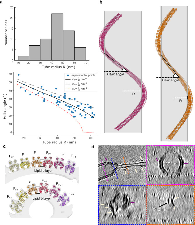
Eps15-homology domain containing proteins (EHDs) are eukaryotic, dynamin-related ATPases involved in cellular membrane trafficking. They oligomerize on membranes into filaments that induce membrane tubulation. While EHD crystal structures in open and closed conformations were previously reported, little structural information is available for the membrane-bound oligomeric form. Consequently, mechanistic insights into the membrane remodeling mechanism have remained sparse. Here, by using cryo-electron tomography and subtomogram averaging, we determined structures of nucleotide-bound EHD4 filaments on membrane tubes of various diameters at an average resolution of 7.6 Å. Assembly of EHD4 is mediated via interfaces in the G-domain and the helical domain. The oligomerized EHD4 structure resembles the closed conformation, where the tips of the helical domains protrude into the membrane. The variation in filament geometry and tube radius suggests a spontaneous filament curvature of approximately 1/70 nm-1. Combining the available structural and functional data, we suggest a model for EHD-mediated membrane remodeling.


Conflict of interest statement

The authors declare no competing interests.

Figures

Fig. 1
Fig. 1. Structure determination of membrane-bound EHD4.
a Tomogram reconstruction of EHD4-covered membrane tubes. The boxed area is magnified in the left bottom corner. Scale bar is 100 nm. b Projection of subtomogram averages of individual tubes with different diameters and helical families. Each tube was individually cropped and averaged using Dynamo. Top: Cross section of a tube. Tubes are ordered from the smallest to biggest diameters. Bottom: Z projection of EHD4 tubes that show different helical families according to different diameters. c Projections of the membrane-bound map using 23,813 particles. d Subtomogram average of the membrane-bound EHD4ΔN complexed with AMPPNP at 7.6 Å resolution. Domain organization of EHD4 in the filaments in two orientations is shown. EH domains (light green), G-domains (orange), helical domains (teal) and the lipid bilayer (white) are colored individually. e Reconstructed EHD4 right-handed helical filaments wrapping around a membrane tube. Each filament is indicated in a different color.
Fig. 2
Fig. 2. Architecture of the EHD4 filament.
a Asymmetric unit of the cryo-ET reconstruction. In the central EHD4 dimer, the domains are colored according to the domain architecture. The cryo-ET density is indicated as grey surface. b The filament is formed by three contact sites. Interface-1 comprises a dimer interface in the G-domain, interface-2 is built by contacts between the G-domain and helical domain whereas interface-3 represents the G-interface and may involve the nucleotide.
Fig. 3
Fig. 3. The membrane-bound EHD4 structure explains the effects of mutations.
Previously reported point mutants of EHD2 and EHD4 were plotted on the EHD4 structure. The effects of the mutations and the relevant literature is listed. All mutagenesis data are consistent with our oligomerized EHD4 structure.
Fig. 4
Fig. 4. The membrane-binding mode of EHD4.
a The cryo-ET model of EHD4 (teal) was superimposed with the G-domain on the crystal structures of EHD2 (yellow; pdb code 4CID) and EHD4 (pink; pdb code 5MTV). The comparison reveals that the membrane-bound structure adopts a closed conformation akin to EHD2. b Membrane-binding site of EHD4 inserts into the lipid outer layer. The two lower panels show how the membrane interaction of the EHD4 filaments induces buckling of the lipid outer layer, as indicated by the magenta dotted line. c Membrane-binding site of EHD4 based on the fittings shown in a and b. Side chains were modelled based on the EHD4 crystal structure. d Sequence alignments of EHD proteins of mouse (mm - Mus musculus) and human (hs – Homo sapiens) reveals a high conservation of the membrane binding site. Conserved residues in all 4 EHD proteins are shown in the sequence alignment as dots (.). Residues that differ in EHDs are highlighted (*).
Fig. 5
Fig. 5. EHD4 filaments adapt to the curvature of membrane tubes.
a Distribution of tube radius (top) and the relation of tube radius and orientation of the filament along the tube axis (bottom). Solid line shows the best fitting spontaneous curvature (see Methods). Dotted lines show the limiting range to illustrate the confidence in predicting κ0. b EHD4-covered membrane tubes show various assemblies. A single filament is depicted around the lipid tube (gray). The average helical assembly of the membrane-bound EHD4 structure is represented on the left and the tube with the smallest diameter is depicted on the right. Both filaments have 42 EHD4 dimers. c EHD4 filaments adopt different conformations in different tube diameters. The angle between filaments increases in tubes with higher curvature (bottom) along interface-4 (boxed) which acts as a hinge. d De-noised tomographic reconstruction of EHD4 oligomer on a thinning membrane. Cross sections along the tube are shown in magenta, blue and orange dashed boxes. Ordered EHD4 filaments form on a tube with a wide diameter (purple arrow), while membrane-bound EHD4 dimers (yellow arrows) were found on a narrow tube. Source data are provided as a Source Data file.
Fig. 6
Fig. 6. Model for EHD-mediated membrane remodeling.
a 1) ATP-bound EHD dimers are recruited to flat membranes in the open conformation where they oligomerize via interface-2 into filaments of low curvature. 2) Membrane curvature induces the transition of the open to closed conformation. In turn, insertion of the membrane-binding site into the membrane promotes membrane curvature, which is associated with the formation of a stable helical filament via interface-3 (see also Supplementary Movies 3 and 4). Further constriction of the membrane tube will lead to an increase of the helical pitch. b ATP hydrolysis is expected to destabilize the G interface, leading to disassembly of the filament. The ADP-bound EHD dimer may convert back to the open conformation and dissociate from the membrane.

References

    1. Naslavsky N, Caplan S. EHD proteins: key conductors of endocytic transport. Trends Cell Biol. 2011;21:122–131. doi: 10.1016/j.tcb.2010.10.003. - DOI - PMC - PubMed
    1. Pohl U, et al. EHD2, EHD3, and EHD4 encode novel members of a highly conserved family of EH domain-containing proteins. Genomics. 2000;63:255–262. doi: 10.1006/geno.1999.6087. - DOI - PubMed
    1. Grant B, et al. Evidence that RME-1, a conserved C. elegans EH-domain protein, functions in endocytic recycling. Nat. Cell Biol. 2001;3:573–579. doi: 10.1038/35078549. - DOI - PubMed
    1. Lin SX, Grant B, Hirsh D, Maxfield FR. Rme-1 regulates the distribution and function of the endocytic recycling compartment in mammalian cells. Nat. Cell Biol. 2001;3:567–572. doi: 10.1038/35078543. - DOI - PubMed
    1. Caplan S, et al. A tubular EHD1-containing compartment involved in the recycling of major histocompatibility complex class I molecules to the plasma membrane. EMBO J. 2002;21:2557–2567. doi: 10.1093/emboj/21.11.2557. - DOI - PMC - PubMed

Publication types

MeSH terms