Review
. 2022 Nov 25;11(23):3768.
doi: 10.3390/cells11233768.

Modulation of the Microglial Nogo-A/NgR Signaling Pathway as a Therapeutic Target for Multiple Sclerosis

Affiliations
Review

Modulation of the Microglial Nogo-A/NgR Signaling Pathway as a Therapeutic Target for Multiple Sclerosis

Danica Nheu et al. Cells. .

Abstract

Current therapeutics targeting chronic phases of multiple sclerosis (MS) are considerably limited in reversing the neural damage resulting from repeated inflammation and demyelination insults in the multi-focal lesions. This inflammation is propagated by the activation of microglia, the endogenous immune cell aiding in the central nervous system homeostasis. Activated microglia may transition into polarized phenotypes; namely, the classically activated proinflammatory phenotype (previously categorized as M1) and the alternatively activated anti-inflammatory phenotype (previously, M2). These transitional microglial phenotypes are dynamic states, existing as a continuum. Shifting microglial polarization to an anti-inflammatory status may be a potential therapeutic strategy that can be harnessed to limit neuroinflammation and further neurodegeneration in MS. Our research has observed that the obstruction of signaling by inhibitory myelin proteins such as myelin-associated inhibitory factor, Nogo-A, with its receptor (NgR), can regulate microglial cell function and activity in pre-clinical animal studies. Our review explores the microglial role and polarization in MS pathology. Additionally, the potential therapeutics of targeting Nogo-A/NgR cellular mechanisms on microglia migration, polarization and phagocytosis for neurorepair in MS and other demyelination diseases will be discussed.

Keywords: Nogo receptor; Nogo-A; alternatively activated microglia; classically activated microglia; microglia; microglial polarization; multiple sclerosis; myelin debris.


Conflict of interest statement

The authors declare no conflict of interest.

Figures
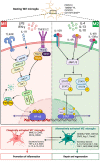
Figure 1
Figure 1
The mechanistic activation of classically activated ‘M1′ and alternatively activated ‘M2′ microglia. In the presence of interferon-gamma (IFN-γ) and lipopolysaccharides (LPS), the resting ‘M0′ microglia undergo polarization towards the classically activated ‘M1′ proinflammatory phenotype by activation of the nuclear factor kappa beta (NF-κB)-dependent pathway. In contrast, the IL-4, IL-10 and IL-13 cytokines trigger signal transducer and activator of transcription (STAT)3/STAT6 phosphorylation within the resting microglia, transitioning cells into the alternatively activated phenotype and increasing the translation of anti-inflammatory cytokines. Under proinflammatory conditions, the classically activated ‘M1′ microglia exacerbate neuroinflammation, whilst the alternatively activated ‘M2′ phenotype can promote repair and regeneration. BDNF: Brain-derived neurotrophic factor. Chil3: Chitinase-like protein 3. CSF-1: Macrophage colony-stimulating factor-1. FGF: Fibroblast growth factors. FIZZ: Found in inflammatory zone. GDNF: Glial cell-derived neurotrophic factor. IGF: Insulin-like growth factor. IKK: I kappa β kinase. IRAK: Interleukin-1 receptor-associated kinase 1. JAK: Janus kinase. MHC: Major histocompatibility complex. MYD88: Myeloid differentiation primary response 88. NEMO: Nuclear kappa- β essential modulator. NGF: Nerve growth factor. RIPK1: Receptor-interacting serine/threonine protein kinase 1. TAB: Transforming growth factor β activated kinase 1 and MAP3K7 binding protein 2. TAK1: Transforming growth factor β activated kinase 1. TGF-β: Transforming growth factor β. TLR: Toll-like receptor. TNF: Tumor necrosis factor. TRADD: Tumor necrosis factor receptor type 1-associated DEATH domain protein. TRAF: Tumor necrosis factor receptor-associated factor. TREM2: Triggering receptor expressed on myeloid cells 2. [Illustration created in BioRender.com accessed on 17 October 2022].
Figure 2
Figure 2
Microglia during chronic neuroinflammatory and neurodegenerative conditions. B cells with other lymphocytes and myeloid cells residing in the cerebrospinal fluid (CSF) infiltrate the subpial and meningeal cerebral cortex and the region of perivascular spaces, forming ectopic B cell follicle-like lesions as observed in progressive multiple sclerosis. As these cells percolate through the cortices towards the axon, resulting in chronic inflammation in grey and white matter, microglia that are activated by bound myelin-specific immunoglobulins secrete proinflammatory cytokines, contributing to promulgated myelin and axonal damage. Under neurodegenerative conditions, the disrupted myelin sheath can act as one of the main precursors for further axonal damage, followed by the formation of reactive astrocytic scarring potentiated through the interaction with activated microglia. [Illustration created in BioRender.com accessed on 17 October 2022].

Similar articles

Cited by

References

    1. Popescu B.F.G., Pirko I., Lucchinetti C.F. Pathology of multiple sclerosis: Where do we stand? Contin. Minneap Minn. 2013;19:901–921. doi: 10.1212/01.CON.0000433291.23091.65. - DOI - PMC - PubMed
    1. Dubey D., Sguigna P., Stüve O. Managing Disability in Progressive Multiple Sclerosis. Curr. Treat. Options Neurol. 2016;18:27. doi: 10.1007/s11940-016-0412-7. - DOI - PubMed
    1. Cadavid D., Cohen J.A., Freedman M.S., Goldman M.D., Hartung H.P., Havrdova E., Jeffery D., Kapoor R., Miller A., Sellebjerg F., et al. The EDSS-Plus, an improved endpoint for disability progression in secondary progressive multiple sclerosis. Mult. Scler. 2017;23:94–105. doi: 10.1177/1352458516638941. - DOI - PubMed
    1. Paolicelli Rosa C., Bolasco G., Pagani F., Maggi L., Scianni M., Panzanelli P., Giustetto M., Ferreira Tiago A., Guiducci E., Dumas L., et al. Synaptic Pruning by Microglia Is Necessary for Normal Brain Development. Science. 2011;333:1456–1458. doi: 10.1126/science.1202529. - DOI - PubMed
    1. Pencea V., Bingaman K.D., Wiegand S.J., Luskin M.B. Infusion of Brain-Derived Neurotrophic Factor into the Lateral Ventricle of the Adult Rat Leads to New Neurons in the Parenchyma of the Striatum, Septum, Thalamus, and Hypothalamus. J. Neurosci. 2001;21:6706. doi: 10.1523/JNEUROSCI.21-17-06706.2001. - DOI - PMC - PubMed

Grants and funding