. 2022 Nov 25;11(23):3774.
doi: 10.3390/cells11233774.

Vascularization of Poly-ε-Caprolactone-Collagen I-Nanofibers with or without Sacrificial Fibers in the Neurotized Arteriovenous Loop Model

Affiliations

Vascularization of Poly-ε-Caprolactone-Collagen I-Nanofibers with or without Sacrificial Fibers in the Neurotized Arteriovenous Loop Model

Simon Kratzer et al. Cells. .

Abstract

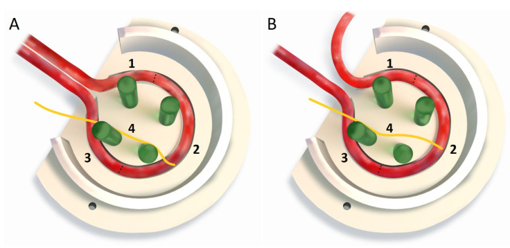
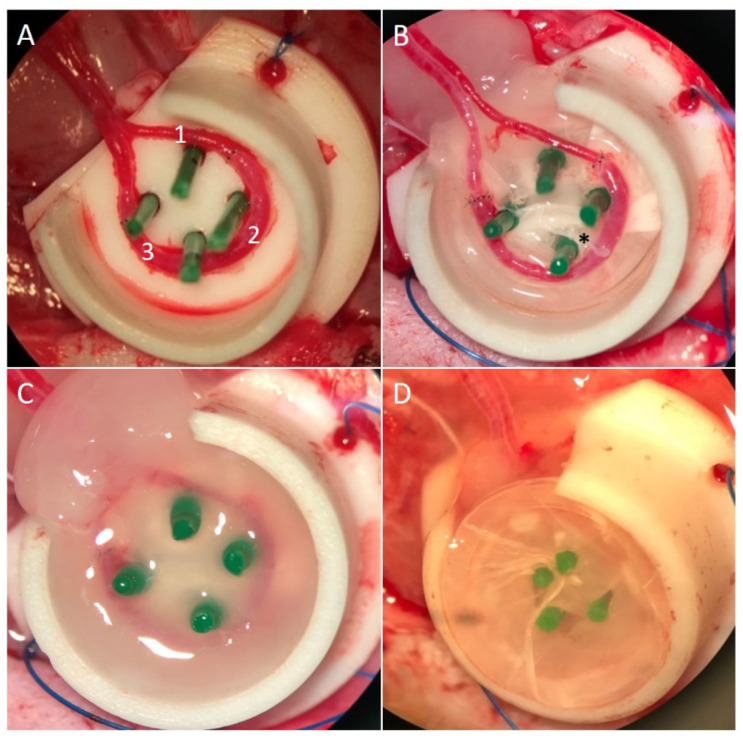
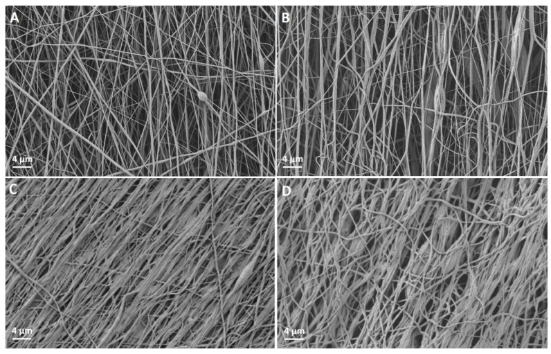
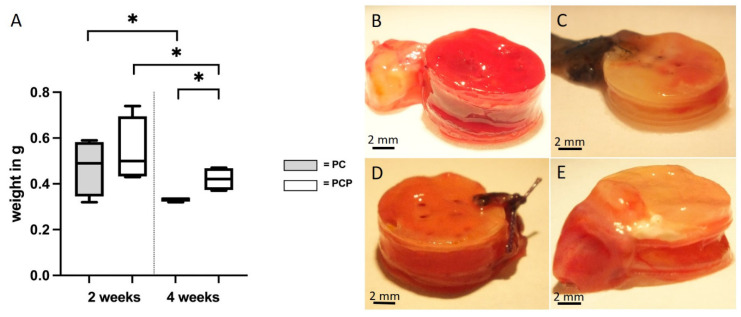
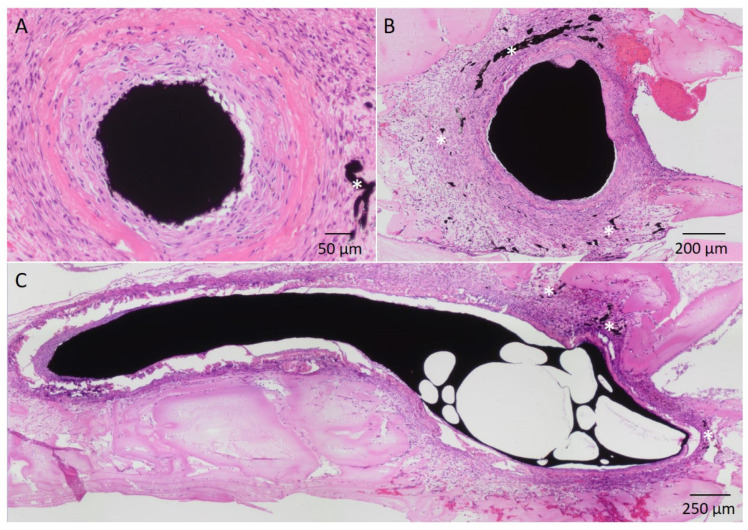
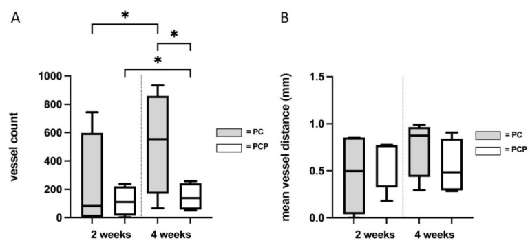
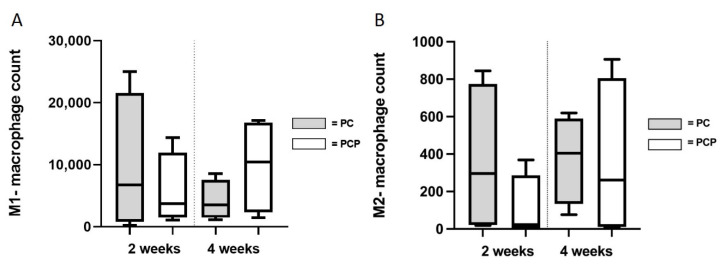
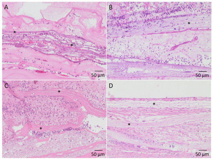
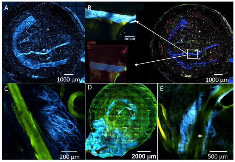
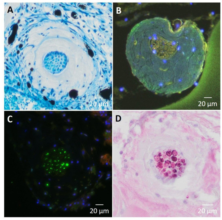
Electrospun nanofibers represent an ideal matrix for the purpose of skeletal muscle tissue engineering due to their highly aligned structure in the nanoscale, mimicking the extracellular matrix of skeletal muscle. However, they often consist of high-density packed fibers, which might impair vascularization. The integration of polyethylene oxide (PEO) sacrificial fibers, which dissolve in water, enables the creation of less dense structures. This study examines potential benefits of poly-ε-caprolactone-collagen I-PEO-nanoscaffolds (PCP) in terms of neovascularization and distribution of newly formed vessels compared to poly-ε-caprolactone -collagen I-nanoscaffolds (PC) in a modified arteriovenous loop model in the rat. For this purpose, the superficial inferior epigastric artery and vein as well as a motor nerve branch were integrated into a multilayer three-dimensional nanofiber scaffold construct, which was enclosed by an isolation chamber. Numbers and spatial distribution of sprouting vessels as well as macrophages were analyzed via immunohistochemistry after two and four weeks of implantation. After four weeks, aligned PC showed a higher number of newly formed vessels, regardless of the compartments formed in PCP by the removal of sacrificial fibers. Both groups showed cell influx and no difference in macrophage invasion. In this study, a model of combined axial vascularization and neurotization of a PCL-collagen I-nanofiber construct could be established for the first time. These results provide a foundation for the in vivo implantation of cells, taking a major step towards the generation of functional skeletal muscle tissue.

Keywords: AV loop model; EPI loop model; PCL-collagen I-nanofiber scaffolds; neoangiogenesis; neurotization; polyethylene oxide; vascularization of nanofiber scaffolds.


Conflict of interest statement

The authors declare no conflict of interest. Any influence of the funding sources on the study design, analysis and interpretation of the results can be excluded by the authors. No benefit of any kind will be received either directly or indirectly by the authors.

Figures

Figure 1
Figure 1
A: Comparison between the newly modified (A) and the previously described (B) EPI loop model, showing the different entry angles of the arteries into the chamber. EPI loop consisting of SIEA (1), vein interposition (2), SIEV (3) and motoric nerve branch (4) B: EPI loop consisting of saphenous artery (1), vein interposition (2), SIEV (3) and motoric nerve branch (4).
Figure 2
Figure 2
(A): EPI loop consisting of SIEA (1), vein interposition (2) and ipsilateral SIEV (3); (B): PTFE chamber including four spacers and two layers of nanofiber scaffolds, EPI loop with obturator nerve (*); (C): Fibrin gel on top of scaffolds and EPI loop; (D): two more layers of PCL-collagen/PCL-collagen-PEO nanofiber scaffolds on top.
Figure 3
Figure 3
(A): PCL-collagen I-nanofibers before pretreatment. (B): PCL-collagen I-PEO-nanofibers before pretreatment. (C): PCL-collagen I-nanofibers after treatment with EtOH and PBS. (D): PCL-collagen I-PEO-nanofibers after treatment with EtOH and PBS.
Figure 4
Figure 4
Explanted scaffold-EPI loop-constructs. (A): Explant weights. Box plots demonstrate decrease from two weeks to four weeks of implantation and differences of PC and PCP after four weeks of implantation (* p ≤ 0.05, unpaired t-test); (B): PCL-collagen I-scaffold-construct after two weeks of implantation (C): PCL-collagen I-scaffold-construct after four weeks of implantation (D): PCL-collagen I-PEO-scaffold-construct after two weeks of implantation (E): PCL-collagen I-PEO-scaffold-construct after four weeks of implantation.
Figure 5
Figure 5
H&E stained sections of a two weeks PCL-collagen I-PEO-construct. Exemplary newly formed vessels are indicated by (*). (A): Superficial inferior epigastric artery filled with India ink; (B): Superficial inferior epigastric vein filled with India ink, surrounded by newly formed vessels. (C): Longitudinal section through the venous interposition filled with India ink, surrounded by newly formed vessels.
Figure 6
Figure 6
(A): Number of newly formed vessels. (B): Mean vessel distance of newly formed vessels to the main EPI loop vessels. An unpaired t-test or Mann–Whitney test was performed as appropriate. Statistically significant differences are marked with * for p ≤ 0.05.
Figure 7
Figure 7
Count of pro- (M1) (A) and anti-inflammatory (M2) (B) macrophages. No differences in the immunological interaction of PCL-collagen I-scaffolds or PCL-collagen I-PEO-scaffolds were found after two weeks or four weeks.
Figure 8
Figure 8
H&E stained sections showing infiltration of cells into the nanofiber scaffold layers. Exemplary nanofibers are marked by (*) (A): PCL-collagen I-PEO-scaffolds after two weeks of implantation; (B): PCL-collagen I-PEO-scaffolds after four weeks of implantation; (C): PCL-collagen I-scaffolds after two weeks of implantation; (D): PCL-collagen I-scaffolds after four weeks of implantation.
Figure 9
Figure 9
Color pattern: Blue indicates the SHG signal of collagen in vascular and nerve structures. Green and red represent the tissue autofluorescence. (A): EPI loop PC construct with subsequent explantation. Strong signal of motor nerve branch and vessels in SHG channel; (B): Positive signal of motor nerve branch in SHG channel; (C): newly formed capillaries (blue) in PC4; (D): Visualization of the EPI loop model (PCP) after four weeks of implantation; (E): Neoangiogenesis (blue) in the area of the EPI loop (indicated by (*)).
Figure 10
Figure 10
(A): Cross section through the motor nerve branch, stained with methylene blue (PCP4). Newly formed vessels filled with India ink show pronounced vascularization in the vicinity of the nerve. (B) (PC2) and (C) (PCP4): Synaptophysin staining of axon cross-section shows the presence of synaptic vesicle proteins p38 (green). (D): S100 immunohistochemistry showing peripheral glial cells (Schwann cells) between the axons of the motor nerve (PCP2).

References

    1. Cai A., Hardt M., Schneider P., Schmid R., Lange C., Dippold D., Schubert D.W., Boos A.M., Weigand A., Arkudas A., et al. Myogenic differentiation of primary myoblasts and mesenchymal stromal cells under serum-free conditions on PCL-collagen I-nanoscaffolds. BMC Biotechnol. 2018;18:75. doi: 10.1186/s12896-018-0482-6. - DOI - PMC - PubMed
    1. Willett N.J., Krishnan L., Li M.T., Guldberg R.E., Warren G.L. Guidelines for Models of Skeletal Muscle Injury and Therapeutic Assessment. Cells Tissues Organs. 2016;202:214–226. doi: 10.1159/000445345. - DOI - PubMed
    1. Horch R.E., Weigand A., Wajant H., Groll J., Boccaccini A.R., Arkudas A. Biofabrication: New approaches for tissue regeneration. Handchir. Mikrochir. Plast. Chir. 2018;50:93–100. doi: 10.1055/s-0043-124674. - DOI - PubMed
    1. Huang R.L., Kobayashi E., Liu K., Li Q. Bone Graft Prefabrication Following the In Vivo Bioreactor Principle. EBioMedicine. 2016;12:43–54. doi: 10.1016/j.ebiom.2016.09.016. - DOI - PMC - PubMed
    1. Robering J.W., Al-Abboodi M., Titzmann A., Horn I., Beier J.P., Horch R.E., Kengelbach-Weigand A., Boos A.M. Tissue Engineering of Lymphatic Vasculature in the Arteriovenous Loop Model of the Rat. Tissue Eng. Part A. 2021;27:129–141. doi: 10.1089/ten.tea.2020.0108. - DOI - PubMed

LinkOut - more resources