. 2022 Nov 26;11(23):3792.
doi: 10.3390/cells11233792.

CircZXDC Promotes Vascular Smooth Muscle Cell Transdifferentiation via Regulating miRNA-125a-3p/ABCC6 in Moyamoya Disease

Affiliations

CircZXDC Promotes Vascular Smooth Muscle Cell Transdifferentiation via Regulating miRNA-125a-3p/ABCC6 in Moyamoya Disease

Yuan Liu et al. Cells. .

Abstract

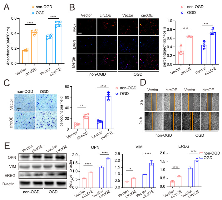
Moyamoya disease (MMD) is an occlusive, chronic cerebrovascular disease affected by genetic mutation and the immune response. Furthermore, vascular smooth muscle cells (VSMCs) and endothelial cells (ECs) participate in the neointima of MMD, but the etiology and pathophysiological changes in MMD vessels remain largely unknown. Therefore, we established the circZXDC (ZXD family zinc finger C)-miR-125a-3p-ABCC6 (ATP-binding cassette subfamily C member 6) axis from public datasets and online tools based on "sponge-like" interaction mechanisms to investigate its possible role in VSMCs. The results from a series of in vitro experiments, such as dual luciferase reporter assays, cell transfection, CCK-8 assays, Transwell assays, and Western blotting, indicate a higher level of circZXDC in the MMD plasma, especially in those MMD patients with the RNF213 mutation. Moreover, circZXDC overexpression results in a VSMC phenotype switching toward a synthetic status, with increased proliferation and migration activity. CircZXDC sponges miR-125a-3p to increase ABCC6 expression, which induces ERS (endoplasmic reticulum stress), and subsequently regulates VSMC transdifferentiation from the contractive phenotype to the synthetic phenotype, contributing to the intima thickness of MMD vessels. Our findings provide insight into the pathophysiological mechanisms of MMD and indicate that the circZXDC-miR-125a-3p-ABCC6 axis plays a pivotal role in the progression of MMD. Furthermore, circZXDC might be a diagnostic biomarker and an ABCC6-specific inhibitor and has the potential to become a promising therapeutic option for MMD.

Keywords: ABCC6; ERS; MMD; circRNA; stroke; transdifferentiation.


Conflict of interest statement

The authors declare that they have no competing interests.

Figures

Figure 1
Figure 1
The construction of the circZXDC–miR-125a-3p–ABCC6 axis and the validation of the plasma level of circZXDC as a diagnostic biomarker of MMD. (A) A schematic diagram of how the circRNA–miRNA–mRNA network was established. (B) Final exact network, including the circRNAs, miRNAs, and mRNAs interacting in MMD. (C) The relative expression level of circ0067130 and circ0004508 in the plasma of the non-MMD and MMD patients using RT-qPCR. (D) The receiver operating characteristic (ROC) curve analysis for the evaluation of the diagnosis ability of circ0067130 under R. The left panel illustrates the ROC curve of the MMD patients regardless of RNF213 mutation. The middle panel indicates the ROC curve of RNF213 WT MMD patients’ plasma circ0067130. The cut-off point is 1.304, and the AUC value is 0.86. The right panel depicts the RNF213 p.4810K mutation in the MMD patient’s plasma circ0067130 ROC curve. The cut-off point is 1.238, and the AUC value is 0.92. (E) The fold change of plasma circ0067130 in MMD patients without the RNF213 p.4810K mutation and MMD RNF213 p.4810K mutation patients compared to non-MMD patients with RT-qPCR. The results of all groups are shown as mean ± SD, and student’s t-test was used to compare expression levels or values among different groups, *** p < 0.001., **** p < 0.0001.
Figure 2
Figure 2
VSMCs contribute to neointima in MMD. (A) The RT-qPCR results of the circZXDC relative expression level in VSMCs, HUVEC, and the 293T cells with transferred SiR-RNF213 and NC. (B) HE results of STA vessels in non-MMD and MMD patients (intima displayed between the yellow lines). Scale bar = 50 µm. The correlation between circ0067130 expression and intima thickness. R2 = 0.6334, and the p value is 0.004. (C) α-SMA immunofluorescence staining in MMD STA vessels. Scale bar = 100 µm (α-SMA, green; DAPI, blue). **** p < 0.0001.
Figure 3
Figure 3
CircZXDC overexpression results in VSMC transdifferentiation towards the synthetic phenotype. The results of the CCK-8 assay (A), ki-67 immunofluorescence staining, scale bar = 100 µm (B), Transwell assay, scale bar = 50 µm (C), and the wound-healing assay (D), measuring the VSMCs transferred with the vector plasmid (Vector) and circZXDC overexpression plasmid (circOE) under non-OGD/OGD conditions; (E) Western blot analysis of the protein expression of OPN, VIM, and EREG in the VSMCs transferred with the vector plasmid (Vector) and the circZXDC overexpression plasmid (circOE) under non-OGD/OGD conditions. The results of all the groups are shown as mean ± SD; two-way ANOVA with Tukey’s post hoc test was used. The significance level was accepted as * p < 0.05, ** p < 0.01, *** p < 0.001, **** p < 0.0001.
Figure 4
Figure 4
The regulating role of the circZXDC–miR-125a-3p axis in VSMC transdifferentiation towards the synthetic phenotype. (A) The RT-qPCR results of miR-125a-3p relative expression in VSMCs transferred with vector plasmid (Vector) and circZXDC overexpression plasmid (circOE). (B) The luciferase assay of the targeted relationship between miR-125a-3p and circZXDC. The results of the CCK-8 assay (C), ki-67 immunofluorescence staining; scale bar = 100 µm (D), Transwell assay; scale bar = 50 µm (E); and Western blotting analysis (F) of VSMCs transferred with vector plasmid (Vector) or circZXDC overexpression plasmid (circOE) in combination with NC mimic or miR-125a-3p mimic (miR) under non-OGD/OGD conditions. Two-way ANOVA and the Tukey’s post hoc test was used. The significance level was accepted as * p < 0.05, **** p < 0.0001, ns = no significance.
Figure 5
Figure 5
The elucidation of miR-125a-3p regulating VSMCs transdifferentiation via sponging ABCC6 mRNA. (A) Western blotting analysis of ABCC6 expression in the VSMCs transferred with the vector plasmid (Vector) or the circZXDC overexpression plasmid (circOE) in combination with the NC mimic or miR-125a-3p mimic (miR) under non-OGD/OGD conditions. (B) The luciferase assay of the targeted relationship between miR-125a-3p and ABCC6. (C) Immunofluorescence staining of the non-MMD and MMD patient’s STA. Scale bar = 50 µm (ABCC6, red; α-SMA, green; DAPI, blue. Intima circled between the yellow lines). The results of the CCK-8 assay (D), ki-67 immunofluorescence staining (E), Transwell assay, scale bar = 50 µm (F); and Western blotting analysis (G) of the VSMCs transferred with NC or ABCC6 siRNA (si) under non-OGD/OGD conditions. The results of all groups are shown as mean ± SD, student’s t-test and two-way ANOVA; Tukey’s post hoc test was used. The significance level was accepted as * p < 0.05, **** p < 0.0001.
Figure 6
Figure 6
Endoplasmic reticulum stress makes a valuable contribution to the process of VSMC trans-differentiation, affected by ABCC6. (A) ERS-related proteins (XBP1s and p-EIF), immunohistochemistry, and immunofluorescence staining of non-MMD and MMD patients’ STA vessels, scale bar = 100 µm. (B) The correlation between intima ABCC6 expression level and intima thickness (r = 0.665, p = 0.0068), the correlation between intima XBP1s expression level and intima thickness (r = 0.611, p = 0.0155), and the correlation between ABCC6 expression level and XBP1s expression level in MMD patients’ intima (r = 0.8389, p < 0.0001), scale bar = 50 µm. (C) XBP1s and GRP78 protein of the VSMCs transferred with NC or ABCC6 siRNA (si) under non-OGD/OGD conditions were detected by Western blotting. The results of the CCK-8 assay (D) and Transwell assay (E) of the VSMCs transferred with the vector plasmid or the ABCC6 overexpression plasmid (OE) together, with or without 4-PBA, under non-OGD/OGD conditions, scale bar = 50 µm. (F) Western blot analysis of the protein expression of OPN and EREG in the VSMCs vector group, ABCC6 overexpression (OE) group, and the ABCC6 overexpression (OE) + 4PBA group under OGD conditions. The results of all groups are shown as mean ± SD. Two-way ANOVA and Tukey’s post hoc test was used. The significance level was accepted as * p < 0.05, ** p < 0.01, *** p < 0.001, **** p < 0.0001.

References

    1. Kim J.S. Moyamoya Disease: Epidemiology, Clinical Features, and Diagnosis. J. Stroke. 2016;18:2–11. doi: 10.5853/jos.2015.01627. - DOI - PMC - PubMed
    1. Sun Y., Zhou G., Feng J., Chen L., Liu G., Wang J., Wang Q., Yu J., Yang X., Yang Z., et al. Incidence and prevalence of moyamoya disease in urban China: A nation wide retrospective cohort study. Stroke Vasc. Neurol. 2021;6:615–623. doi: 10.1136/svn-2021-000909. - DOI - PMC - PubMed
    1. Zhang Q., Liu Y., Zhang D., Wang R., Zhang Y., Wang S., Yu L., Lu C., Liu F., Zhou J., et al. RNF213 as the major susceptibility gene for Chinese patients with moyamoya disease and its clinical relevance. J. Neurosurg. 2017;126:1106–1113. doi: 10.3171/2016.2.JNS152173. - DOI - PubMed
    1. Kamada F., Aoki Y., Narisawa A., Abe Y., Komatsuzaki S., Kikuchi A., Kanno J., Niihori T., Ono M., Ishii N., et al. A genome-wide association study identifies RNF213 as the first Moyamoya disease gene. J. Hum. Genet. 2011;56:34–40. doi: 10.1038/jhg.2010.132. - DOI - PubMed
    1. Liu W., Hitomi T., Kobayashi H., Harada K.H., Koizumi A. Distribution of moyamoya disease susceptibility polymorphism p.R4810K in RNF213 in East and Southeast Asian populations. Neurol. Med.-Chir. 2012;52:299–303. doi: 10.2176/nmc.52.299. - DOI - PubMed

MeSH terms

LinkOut - more resources