. 2022 Nov 27;11(23):3797.
doi: 10.3390/cells11233797.

Long-Term Characteristics of Human-Derived Biliary Organoids under a Single Continuous Culture Condition

Affiliations

Long-Term Characteristics of Human-Derived Biliary Organoids under a Single Continuous Culture Condition

Ranan G Aktas et al. Cells. .

Abstract

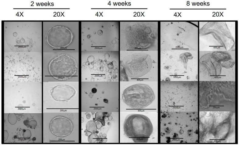
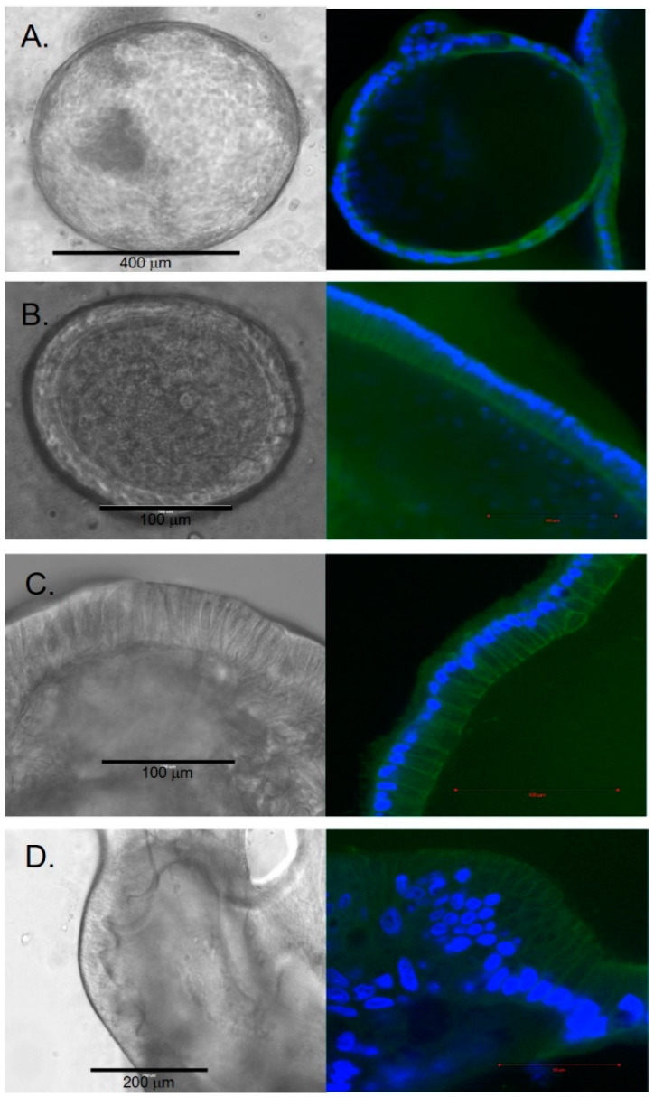
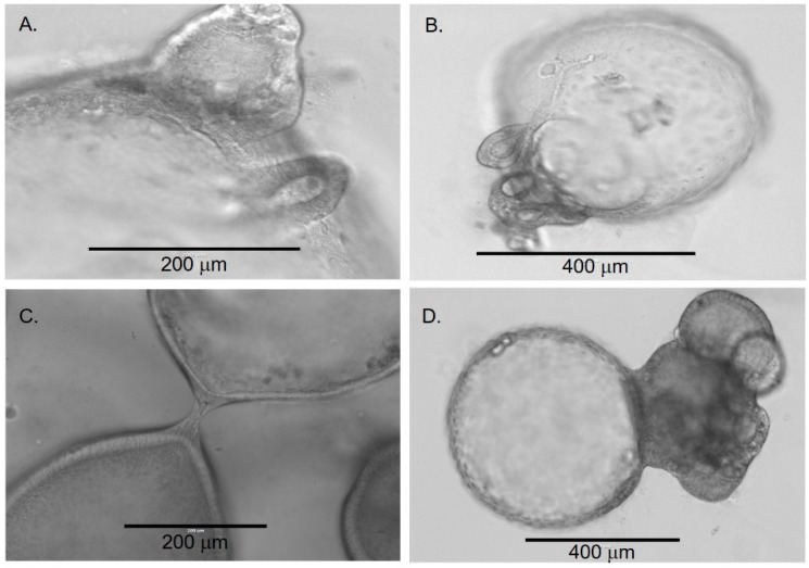
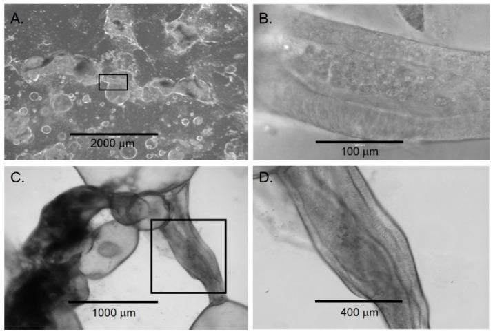
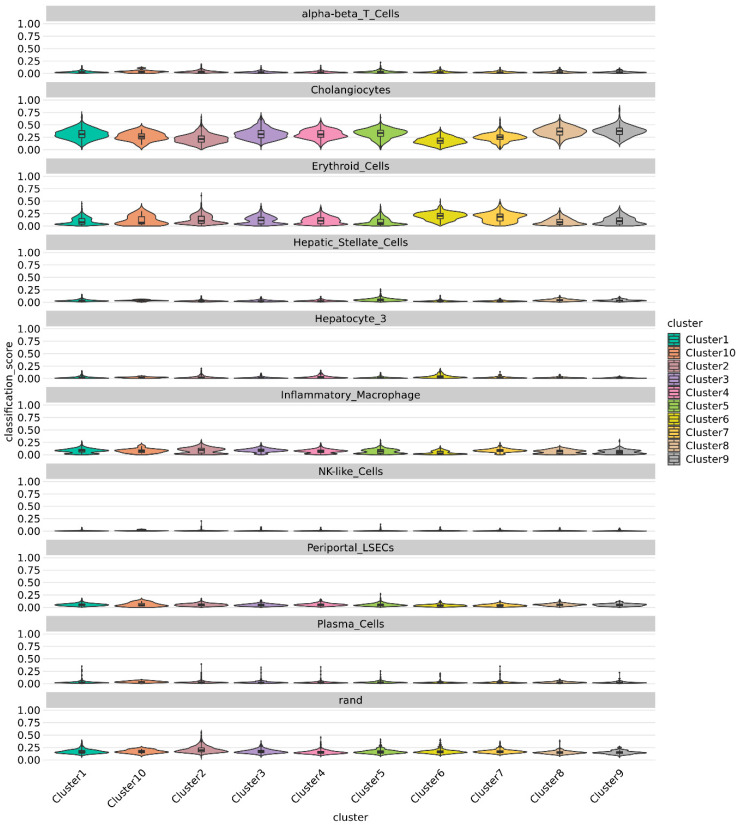
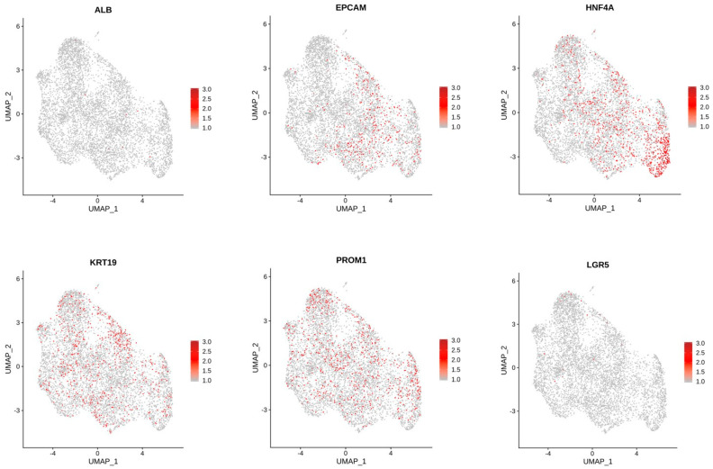
Organoids have been used to investigate the three-dimensional (3D) organization and function of their respective organs. These self-organizing 3D structures offer a distinct advantage over traditional two-dimensional (2D) culture techniques by creating a more physiologically relevant milieu to study complex biological systems. The goal of this study was to determine the feasibility of establishing organoids from various pediatric liver diseases and characterize the long-term evolution of cholangiocyte organoids (chol-orgs) under a single continuous culture condition. We established chol-orgs from 10 different liver conditions and characterized their multicellular organization into complex epithelial structures through budding, merging, and lumen formation. Immunofluorescent staining, electron microscopy, and single-nucleus RNA (snRNA-seq) sequencing confirmed the cholangiocytic nature of the chol-orgs. There were significant cell population differences in the transcript profiles of two-dimensional and organoid cultures based on snRNA-seq. Our study provides an approach for the generation and long-term maintenance of chol-orgs from various pediatric liver diseases under a single continuous culture condition.

Keywords: 3D; biliary system; extracellular matrix; microenvironment; organoid; primary cells; three dimensional.


Conflict of interest statement

The authors declare no conflict of interest.

Figures

Figure 1
Figure 1
Chol-org morphology over 8 weeks from 4 different patient samples with underlying liver disease (from top to bottom: PFICIII, autoimmune hepatitis, alpha-1 antitrypsin deficiency, and ornithine transcarbamylase deficiency). At 2 weeks, chol-orgs demonstrate a simple spheroidal shape which develops into multi-layered and complex structure by 8 weeks.
Figure 2
Figure 2
Cellular histology of chol-orgs with left column representing bright-field images and right column representing immunostaining with CK17 (green) and DAPI (blue) (A) Squamous epithelial morphology is present in early chol-orgs. Over time, cells develop a columnar epithelial morphology (B,C). In late organoids, multi-layered epithelial structures are observed (D).
Figure 3
Figure 3
Transition of early chol-orgs into more complex forms. Budding (A,B) and merging (C,D) processes are observed as the chol-orgs transform into complex structures.
Figure 4
Figure 4
Lumen formation in late chol-orgs. (A) Low-magnification view demonstrates a culture with simple and complex chol-orgs and duct-like structure (black box) which is shown at higher magnification in panel (B). (C) Duct-like structure (black box) between two organoids with higher magnification in panel (D). Note the columnar epithelia along both lateral aspects of the lumen.
Figure 5
Figure 5
Compartment formation in late chol-orgs. (A) Phase contrast image demonstrate several compartments within a late chol-org. (B) IMMUNOFLUORESCENT imaging labeling CFTR.
Figure 6
Figure 6
Immunofluorescence staining of early and late chol-orgs. EpCAM, CFTR, and CK19 membranous staining is present in both early and late organoids. YAP staining is predominantly nuclear in early organoids with increasing cytoplasmic staining in late organoids. Sox9 nuclear staining is patchy and weak in both early and late organoids. Membanous β-catenin staining is present in both early and late organoids. DAPI (blue) is used as a nuclear stain. Scale bars (white line): 100 μm (EpCAM and CFTR); 50 μm (YAP); and 200 μm (Sox9).
Figure 7
Figure 7
Electron microscopy of chol-orgs. (A) Luminal (top of panel) and basal surfaces (bottom of panel) are represented. The pattern of cell nuclei near the basal surface is consistent with a multilayer epithelial structure. (B) Higher magnification image demonstrates concentration of secretory vesicles (denoted by *) near the luminal border of the chol-org. (C) Cilia (arrows) are present along the luminal surface.
Figure 8
Figure 8
Unannotated UMAP clusters from snRNA-seq. (A) Integrated UMAP showing numbers of clusters detected in the snRNA-seq raw data, including both 2D and 3D cells. (B) Comparative UMAP plots showing differences in cell clustering patterns from snRNA-seq data based on 2D or 3D culture conditions. Cluster 2 cell population is significantly decreased in 2D chol-org culture whereas Clusters 5, 8, and 9 constitute a higher proportion of cells in 2D chol-org culture.
Figure 9
Figure 9
Violin plot showing prediction from singleCellNet for each cluster based on top 50 discriminant genes and top 100 gene pairs derived from cell types from MacParland [22] dataset classifier model. All Clusters demonstrate a strong cholangiocytic signature.
Figure 10
Figure 10
Feature Plots showing expression of genes of interest across cells in all clusters.

Similar articles

Cited by

References

    1. Sampaziotis F., de Brito M.C., Madrigal P., Bertero A., Saeb-Parsy K., Soares F.A.C., Schrumpf E., Melum E., Karlsen T.H., Bradley J.A., et al. Cholangiocytes derived from human induced pluripotent stem cells for disease modeling and drug validation. Nat. Biotechnol. 2015;33:845–852. doi: 10.1038/nbt.3275. - DOI - PMC - PubMed
    1. Nuciforo S., Heim M.H. Organoids to model liver disease. JHEP Rep. 2021;3:100198. doi: 10.1016/j.jhepr.2020.100198. - DOI - PMC - PubMed
    1. Sampaziotis F., Muraro D., Tysoe O.C., Sawiak S., Beach T.E., Godfrey E.M., Upponi S.S., Brevini T., Wesley B.T., Garcia-Bernardo J., et al. Cholangiocyte organoids can repair bile ducts after transplantation in the human liver. Science. 2021;371:839–846. doi: 10.1126/science.aaz6964. - DOI - PMC - PubMed
    1. Shiota J., Samuelson L.C., Razumilava N. Hepatobiliary Organoids and Their Applications for Studies of Liver Health and Disease: Are We There Yet? Hepatology. 2021;74:2251–2263. doi: 10.1002/hep.31772. - DOI - PMC - PubMed
    1. Nii T., Makino K., Tabata Y. Three-Dimensional Culture System of Cancer Cells Combined with Biomaterials for Drug Screening. Cancers (Basel) 2020;12:2754. doi: 10.3390/cancers12102754. - DOI - PMC - PubMed

LinkOut - more resources