. 2022 Nov 28;11(23):3804.
doi: 10.3390/cells11233804.

IL-5 and GM-CSF, but Not IL-3, Promote the Proliferative Properties of Inflammatory-like and Lung Resident-like Eosinophils in the Blood of Asthma Patients

Affiliations

IL-5 and GM-CSF, but Not IL-3, Promote the Proliferative Properties of Inflammatory-like and Lung Resident-like Eosinophils in the Blood of Asthma Patients

Jolita Palacionyte et al. Cells. .

Abstract

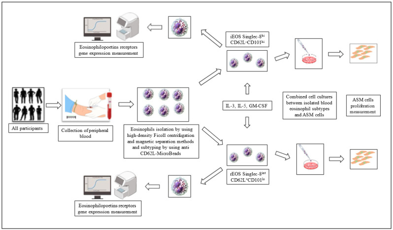
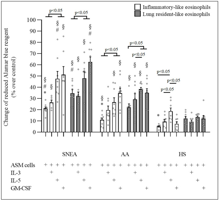
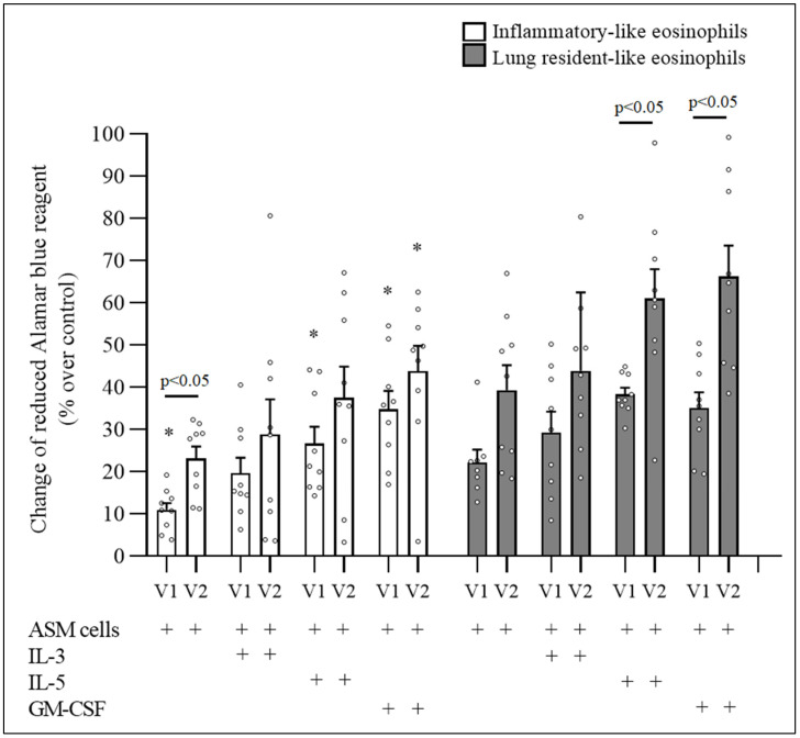
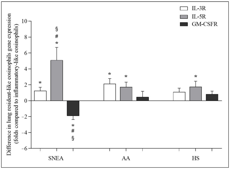
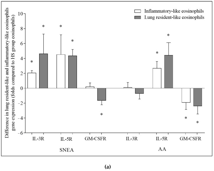
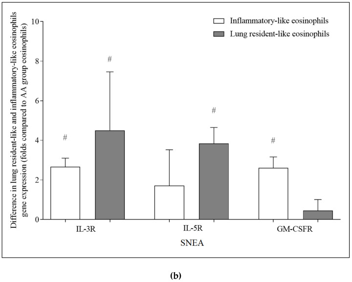
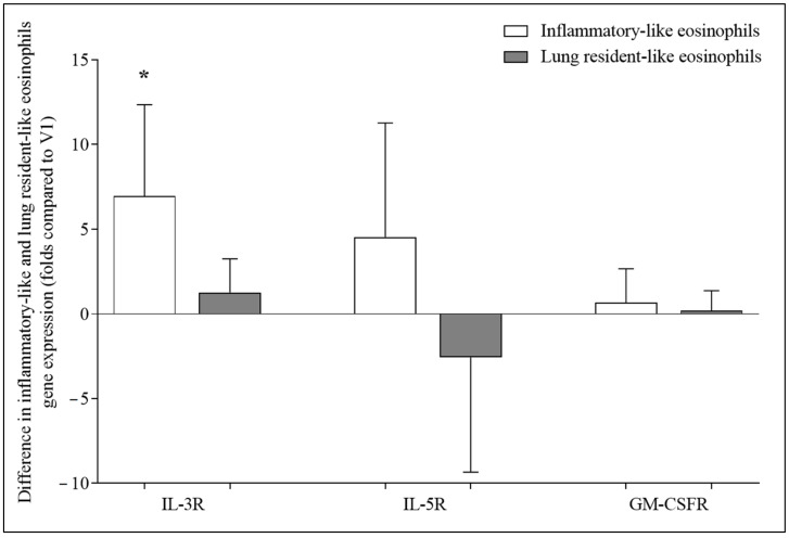
Blood eosinophils can be described as inflammatory-like (iEOS-like) and lung-resident-like (rEOS-like) eosinophils. This study is based on the hypothesis that eosinophilopoetins such as interleukin (IL)-3 and IL-5 and granulocyte-macrophage colony-stimulating factor (GM-CSF) alter the proliferative properties of eosinophil subtypes and may be associated with the expression of their receptors on eosinophils. We investigated 8 individuals with severe nonallergic eosinophilic asthma (SNEA), 17 nonsevere allergic asthma (AA), and 11 healthy subjects (HS). For AA patients, a bronchial allergen challenge with Dermatophagoides pteronyssinus was performed. Eosinophils were isolated from peripheral blood using high-density centrifugation and magnetic separation methods. The subtyping of eosinophils was based on magnetic bead-conjugated antibodies against L-selectin. Preactivation by eosinophilopoetins was performed by incubating eosinophil subtypes with IL-3, IL-5, and GM-CSF, and individual combined cell cultures were prepared with airway smooth muscle (ASM) cells. ASM cell proliferation was assessed using an Alamar blue assay. The gene expression of eosinophilopoetin receptors was analyzed with a qPCR. IL-5 and GM-CSF significantly enhanced the proliferative properties of iEOS-like and rEOS-like cells on ASM cells in both SNEA and AA groups compared with eosinophils not activated by cytokines (p < 0.05). Moreover, rEOS-like cells demonstrated a higher gene expression of the IL-3 and IL-5 receptors compared with iEOS-like cells in the SNEA and AA groups (p < 0.05). In conclusion: IL-5 and GM-CSF promote the proliferative properties of iEOS-like and rEOS-like eosinophils; however, the effect of only IL-5 may be related to the expression of its receptors in asthma patients.

Keywords: GM-CSF; IL-3; IL-5; airway smooth muscle cells; asthma; eosinophil; gene expression; proliferation.


Conflict of interest statement

The authors declare no conflict of interest.

Figures

Figure 1
Figure 1
Graphical overview of the study design. ASM—airway smooth muscle; IL—interleukin; GM-CSF—granulocyte-macrophage colony-stimulating factor; CD—cluster of differentiation.
Figure 2
Figure 2
Effect of eosinophilopoetins on proliferative properties of blood eosinophil subtypes. ASM—airway smooth muscle; IL—interleukin; GM-CSF—granulocyte-macrophage colony-stimulating factor. Severe nonallergic eosinophilic asthma (SNEA) group n = 10, nonsevere allergic asthma (AA) group n = 9, healthy subjects (HS) group n = 8. Data presented as the mean ± standard error of the mean. * p < 0.05 compared with appropriate rEOS-like cells in the same group. # p < 0.05 compared with appropriate eosinophil subtypes from AA group. § p < 0.005 compared with appropriate eosinophil subtypes from HS group. Statistical analysis: between investigated groups, Mann–Whitney two-sided U-test; within one study group, Wilcoxon matched-pair signed-rank two-sided test. Data presented as the mean ± standard error of the mean.
Figure 3
Figure 3
The effect of bronchial allergen challenge on proliferative properties of blood eosinophil subtypes in allergic asthma. ASM—airway smooth muscle; IL—interleukin; GM-CSF—granulocyte-macrophage colony-stimulating factor, V1—visit 1 (before bronchial allergen challenge); V2—visit 2 (24 h after bronchial allergen challenge). Results from independent experiments of AA, n = 8. Data presented as the mean ± standard error of the mean. * p < 0.05 compared with appropriate rEOS-like cells. Statistical analysis: between investigated groups, Mann–Whitney two-sided U-test; within one study group, Wilcoxon matched-pair signed-rank two-sided test. Data presented as the mean ± standard error of the mean.
Figure 4
Figure 4
Differences in expression of IL-3R, IL-5R, and GM-CSFR genes between lung resident-like and inflammatory-like eosinophils in the same groups. IL-3R—interleukin 3 receptor; IL-5R—interleukin 5 receptor; GM-CSFR—granulocyte-macrophage colony-stimulating factor receptor. Severe nonallergic eosinophilic asthma (SNEA) group n = 10, nonsevere allergic asthma (AA) group n = 8, healthy subjects (HS) group n = 11. Data presented as the mean ± standard error of the mean, fold change over expression of IL-3R, IL-5R, and GM-CSFR gene on inflammatory-like eosinophils. * p < 0.05 compared with inflammatory-like eosinophils; # p < 0.05 compared with AA group; § p < 0.05 compared with HS group. Statistical analysis: between investigated groups, Mann–Whitney two-sided U-test; within one study group, Wilcoxon matched-pair signed-rank two-sided test. Data presented as the mean ± standard error of the mean.
Figure 5
Figure 5
Differences in expression of IL-3R, IL-5R, and GM-CSFR genes between eosinophil subtypes. Comparison of (a) SNEA and AA groups with HS group; (b) SNEA group with AA group. IL-3R—interleukin 3 receptor; IL-5R—interleukin 5 receptor; GM-CSFR—granulocyte-macrophage colony-stimulating factor receptor. Severe nonallergic eosinophilic asthma (SNEA) group n = 10, nonsevere allergic asthma (AA) group n = 8, healthy subjects (HS) group n = 11. Data presented as the mean ± standard error of the mean, fold change over expression of IL-3R, IL-5R, and GM-CSFR gene in HS and AA groups. * p < 0.05 compared with HS group, # p < 0.05 compared with AA group. Statistical analysis: between investigated groups, Mann–Whitney two-sided U-test; within one study group, Wilcoxon matched-pair signed-rank two-sided test. Data presented as the mean ± standard error of the mean.
Figure 5
Figure 5
Differences in expression of IL-3R, IL-5R, and GM-CSFR genes between eosinophil subtypes. Comparison of (a) SNEA and AA groups with HS group; (b) SNEA group with AA group. IL-3R—interleukin 3 receptor; IL-5R—interleukin 5 receptor; GM-CSFR—granulocyte-macrophage colony-stimulating factor receptor. Severe nonallergic eosinophilic asthma (SNEA) group n = 10, nonsevere allergic asthma (AA) group n = 8, healthy subjects (HS) group n = 11. Data presented as the mean ± standard error of the mean, fold change over expression of IL-3R, IL-5R, and GM-CSFR gene in HS and AA groups. * p < 0.05 compared with HS group, # p < 0.05 compared with AA group. Statistical analysis: between investigated groups, Mann–Whitney two-sided U-test; within one study group, Wilcoxon matched-pair signed-rank two-sided test. Data presented as the mean ± standard error of the mean.
Figure 6
Figure 6
Differences in the expression of IL-3R, IL-5R, and GM-CSFR genes before and after bronchial allergen challenge. V2, visit 2 (24 h after bronchial allergen challenge). IL-3R—interleukin 3 receptor; IL-5R—interleukin 5 receptor; GM-CSFR—granulocyte-macrophage colony-stimulating factor receptor. Results from independent experiments of AA, n = 8. Data presented as the mean ± standard error of the mean, fold change over expression of IL-3R, IL-5R, and GM-CSFR gene after bronchial allergen challenge. * p < 0.05 compared with V2. Statistical analysis: between investigated groups, Mann–Whitney two-sided U-test; within one study group, Wilcoxon matched-pair signed-rank two-sided test. Data presented as the mean ± standard error of the mean.

Similar articles

Cited by

References

    1. Weller P.F., Spencer L.A. Functions of tissue-resident eosinophils. Nat. Rev. Immunol. 2017;17:746–760. doi: 10.1038/nri.2017.95. - DOI - PMC - PubMed
    1. Park Y.M., Bochner B.S. Eosinophil survival and apoptosis in health and disease. Allergy Asthma Immunol. Res. 2010;2:87–101. doi: 10.4168/aair.2010.2.2.87. - DOI - PMC - PubMed
    1. Kita H. Eosinophils: Multifunctional and distinctive properties. Int. Arch. Allergy Immunol. 2013;161((Suppl. S2)):3–9. doi: 10.1159/000350662. - DOI - PMC - PubMed
    1. Wechsler M.E., Munitz A., Ackerman S.J., Drake M.G., Jackson D.J., Wardlaw A.J., Dougan S.K., Berdnikovs S., Schleich F., Matucci A., et al. Eosinophils in Health and Disease: A State-of-the-Art Review. Mayo. Clin. Proc. 2021;96:2694–2707. doi: 10.1016/j.mayocp.2021.04.025. - DOI - PubMed
    1. Mesni L.C., Raulier S., Paulissen G., Xiao X., Birrell M.A., Pirottin D., Janss T., Starkl P., Ramery E., Henket M., et al. Lung-resident eosinophils represent a distinct regulatory eosinophil subset. J. Clin. Investig. 2016;126:3279–3295. doi: 10.1172/JCI85664. - DOI - PMC - PubMed

Substances