. 2022 Nov 28;11(23):3806.
doi: 10.3390/cells11233806.

The Revelation of Continuously Organized, Co-Overexpressed Protein-Coding Genes with Roles in Cellular Communications in Breast Cancer

Affiliations

The Revelation of Continuously Organized, Co-Overexpressed Protein-Coding Genes with Roles in Cellular Communications in Breast Cancer

Aswathy Mary Paul et al. Cells. .

Abstract

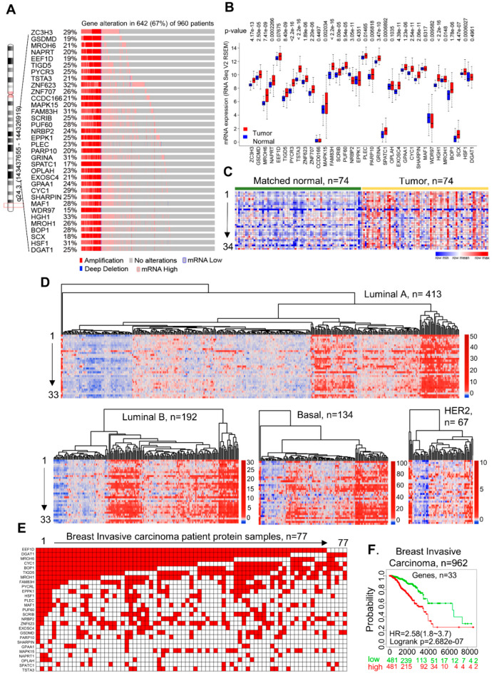
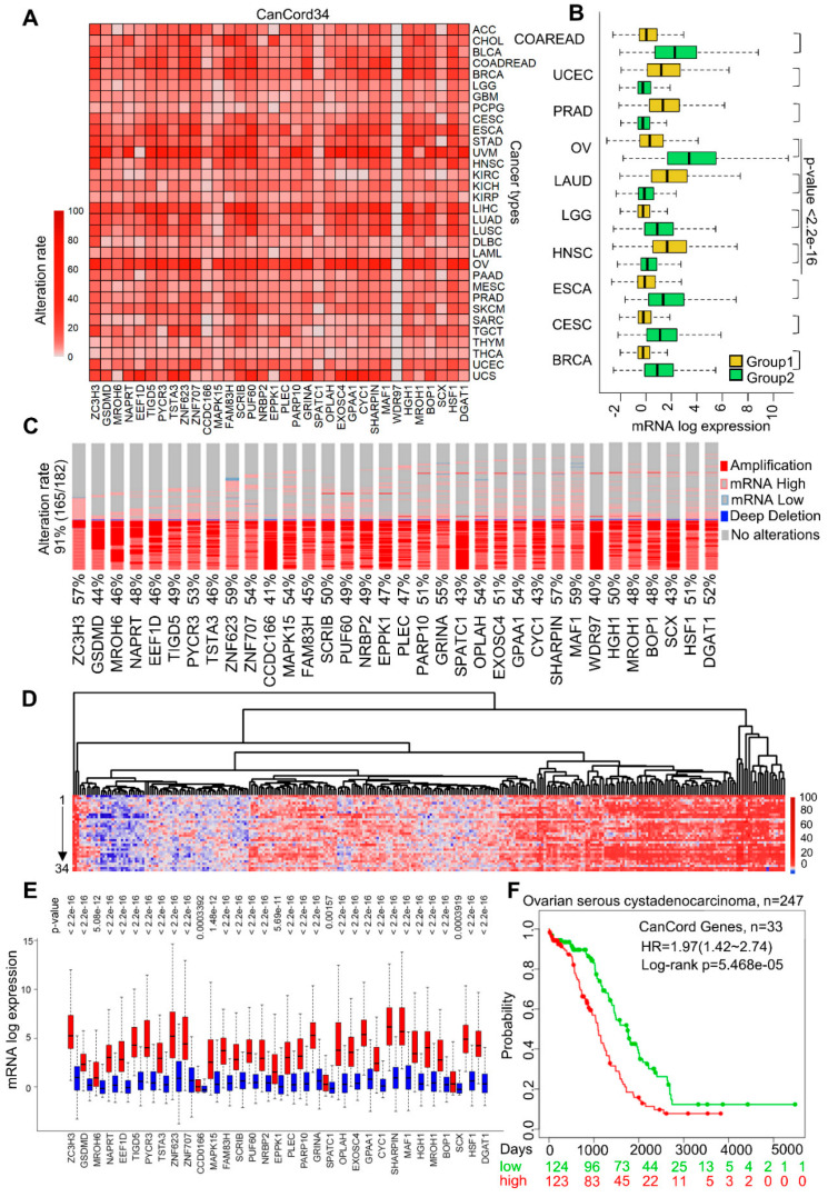
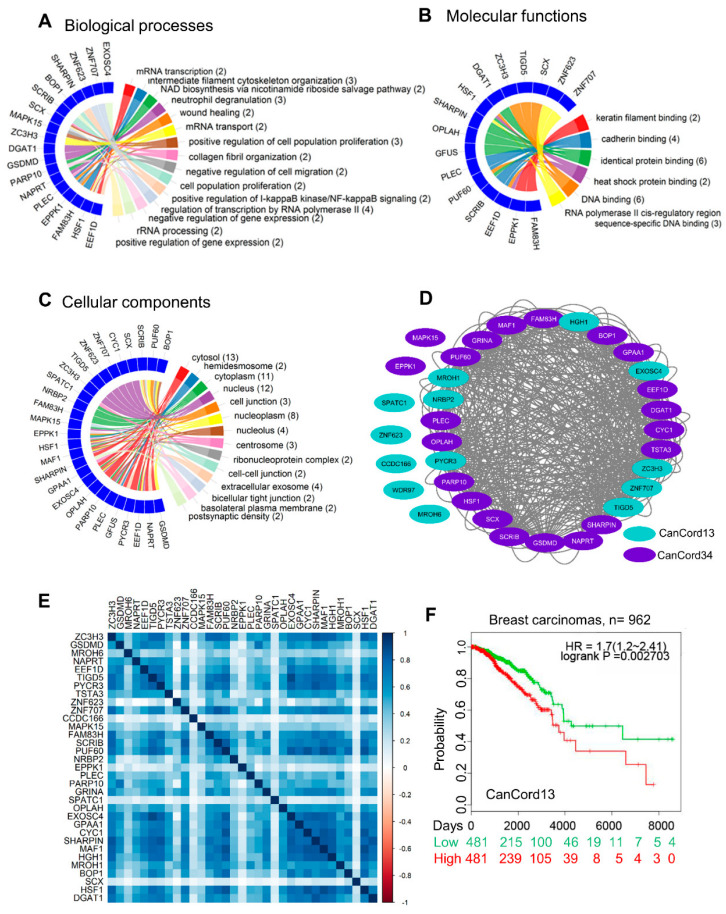
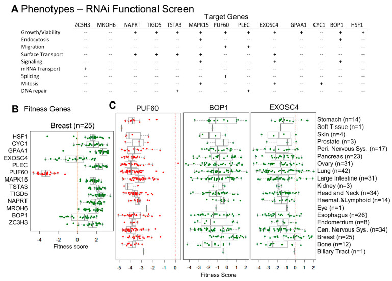
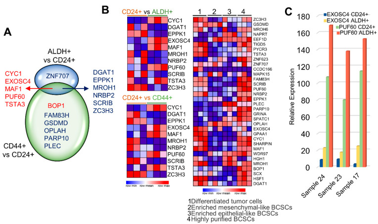
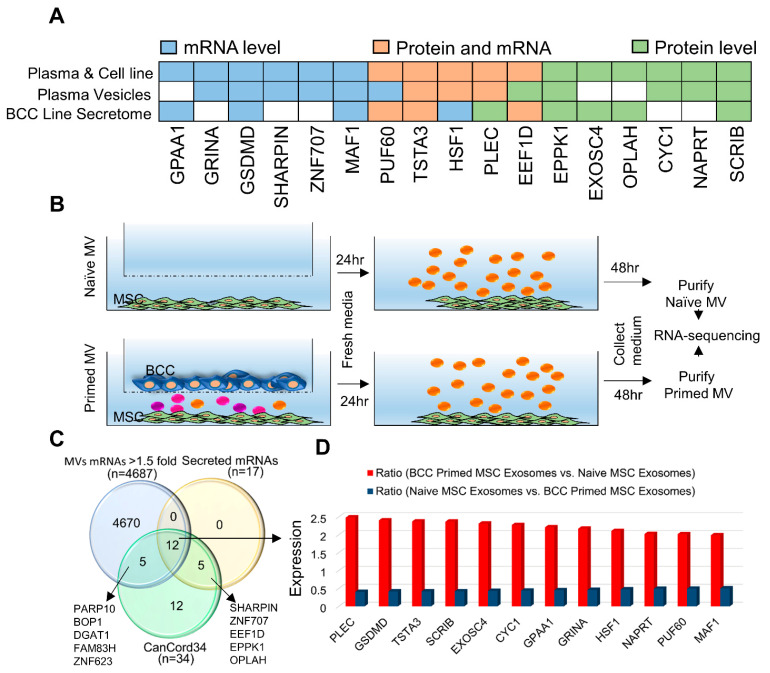
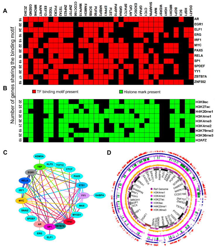
Many human cancers, including breast cancer, are polygenic and involve the co-dysregulation of multiple regulatory molecules and pathways. Though the overexpression of genes and amplified chromosomal regions have been closely linked in breast cancer, the notion of the co-upregulation of genes at a single locus remains poorly described. Here, we describe the co-overexpression of 34 continuously organized protein-coding genes with diverse functions at 8q.24.3(143437655-144326919) in breast and other cancer types, the CanCord34 genes. In total, 10 out of 34 genes have not been reported to be overexpressed in breast cancer. Interestingly, the overexpression of CanCord34 genes is not necessarily associated with genomic amplification and is independent of hormonal or HER2 status in breast cancer. CanCord34 genes exhibit diverse known and predicted functions, including enzymatic activities, cell viability, multipotency, cancer stem cells, and secretory activities, including extracellular vesicles. The co-overexpression of 33 of the CanCord34 genes in a multivariant analysis was correlated with poor survival among patients with breast cancer. The analysis of the genome-wide RNAi functional screening, cell dependency fitness, and breast cancer stem cell databases indicated that three diverse overexpressed CanCord34 genes, including a component of spliceosome PUF60, a component of exosome complex EXOSC4, and a ribosomal biogenesis factor BOP1, shared roles in cell viability, cell fitness, and stem cell phenotypes. In addition, 17 of the CanCord34 genes were found in the microvesicles (MVs) secreted from the mesenchymal stem cells that were primed with MDA-MB-231 breast cancer cells. Since these MVs were important in the chemoresistance and dedifferentiation of breast cancer cells into cancer stem cells, these findings highlight the significance of the CanCord34 genes in cellular communications. In brief, the persistent co-overexpression of CanCord34 genes with diverse functions can lead to the dysregulation of complementary functions in breast cancer. In brief, the present study provides new insights into the polygenic nature of breast cancer and opens new research avenues for basic, preclinical, and therapeutic studies in human cancer.

Keywords: breast cancer; cancer progression; computational biology; coregulation; emerging pathways; polygenic nature; shared mechanisms of gene expression; transcriptome.


Conflict of interest statement

The authors declare no conflict of interest.

Figures

Figure 1
Figure 1
Overview of CanCord34 genes in breast cancer. (A) Dispersal of the genomic alterations in CanCord34 genes in breast cancer patients. Each row represents a gene, and each vertical line represents one patient sample. The alterations are defined with different colors and patterns. The data were taken from the cBioPortal platform. (B) Boxplot image showing differences between the expression patterns of CanCord34 genes in normal and matched tumor samples. The significance of the difference between the gene expression levels was evaluated using the Mann–Whitney–Wilcoxon test. (C) Expression of CanCord34 genes in normal and matched tumor samples. Except for CCDC166, all other genes were upregulated in the tumor samples compared to the normal samples. (D) Heatmap representation of the levels of the CanCord34 mRNAs using the heatmap algorithm of R for different subtypes. The samples with high expression, ~32% of the total samples, are clustered together. Many specimens belonging to the basal and luminal B (LumB) sub-types exhibited the upregulation of CanCord34 genes. (E) Heatmap representation of samples with genes upregulated at the protein level. Each row represents a gene, and the columns represent the tumor samples. Genes are sorted in descending order of gene expression in the tumor samples. (F) Kaplan–Meier survival plots of the CanCord34 genes. Out of 34 genes, 33 were taken to classify the samples, because one of the genes, the CCDC166, could not be mapped in the dataset. The survival plot for the CanCord34 genes was generated using the SurvExpress program.
Figure 2
Figure 2
All-encompassing pattern of CanCord34. (A) A single-color heatmap represents the alteration rate of CanCord34 genes in 33 different cancer types. The increasing intensity of the color gradient is directly proportional to the increasing alteration rate. The X-axis represents the genes, and the Y-axis represents the TCGA abbreviation for the 33 cancer types. (B) Boxplot representation of the expression distribution of CanCord34 genes at the mRNA level for different cancers. Each cancer type was classified into two distinct groups based on CanCord34 expression. (C) Dispersal of alterations in the CanCord34 genes in ovarian cancer patients. Each row represents a gene, and each vertical line represents each patient sample. These genes are coordinately upregulated in patients at the mRNA level. The alterations are represented with different color codes and patterns. The data were taken from cBioPortal. (D) Heatmap representation of CanCord34 gene expression at the mRNA level, constructed using the heatmap algorithm of R for the ovarian cancer data. (E) Boxplot representation of the expression distribution between two groups identified using k-means clustering for each gene. The significance of the noticed difference between the gene expression levels was evaluated using the Mann–Whitney–Wilcoxon test. (F) Kaplan–Meier survival plot for the CanCord34 genes generated using the SurvExpress program.
Figure 3
Figure 3
Functional enrichment analysis of CanCord34 genes. (A) Biological processes. (B) Molecular function. (C) Cellular components. Gene ontology enrichment analyses were performed using the NeVOmics functional annotation program and represented using Circos plots. (D) Circular network representing the CanCord34 inter-correlation. Node colors represent the class of genes as CanCord13 and CanCord34. (E) Functional protein–protein interaction using the online STRING platform. (F) Kaplan–Meier survival analysis of CanCord13 genes using the SurvExpress program.
Figure 4
Figure 4
Towards the functions of CanCord34 genes. (A) Phenotypic analyses of a subset of CanCord34 genes using the functional RNAi screening database. (B) Distribution of a subset of CanCord34 genes as a cellular target with a significant fitness dependency effect upon its knockdown in the test breast cancer cell line. (C) Distribution of a subset of CanCord34 genes as a cellular target with a significant fitness dependency effect upon its knockdown in different cancer cell lines.
Figure 5
Figure 5
CanCord34 genes in cancer stem cells. (A) Venn diagram representing the CanCord34 genes reported to be involved in the stem cells. Upregulated genes are represented in red, whereas downregulated genes are in blue. (B) Heatmap representation of the CanCord34 gene expression in ALDH+ vs. CD24+ cell clusters (top left), CD24+ vs. CD44+ cells, and differentiated tumor cells, enriched mesenchymal-like BCSCs, enriched epithelial-like BCSCs, and highly purified BCSCs. The image was constructed using the heatmap package of Gene Patterns. (C) Bar chart representing the expressions of EXOSC4 and PUF60 in the sample sets of ALDH+ and CD24+ cells.
Figure 6
Figure 6
CanCord34 in secretory functions. (A) The literature curation of CanCord34 genes shows a subset of CanCord34 genes as secreted mRNA, proteins, or both. (B) Cartoon showing the model system used. (C) Venn diagram showing the overlap of the 12 upregulated mRNAs present in MSC-derived extracellular vesicles (n = 4686, >1.5-fold with a p-value 0.05), secreted CanCord34 mRNAs and/or proteins identified in public databases (n = 17), and CanCord34 genes (n = 34). (D) Bar chart showing the upregulation of 12 secreted CanCord34 genes in MSCs incubated with primed exosomes vs. naïve MSC exosomes.
Figure 7
Figure 7
Shared regulatory elements of CanCord34 genes. (A) Shared transcription factors associated with the CanCord34 genes within 1 kb from the TSS. (B) A cluster matrix showed the activating histone marks shared by the CanCord34 genes in MCF-7 cells. (C) Interactions and heterodimerization of TFs with the base TFs (in which interacting response elements are present) in CanCord34 genes. (D) Histone mark enrichment analysis of CanCord34 genes and the status of activated histone marks, represented as a Circos plot.

Similar articles

Cited by

References

    1. Horne S.D., Pollick S.A., Heng H.H.Q. Evolutionary Mechanism Unifies the Hallmarks of Cancer. Int. J. Cancer. 2015;136:2012–2021. doi: 10.1002/ijc.29031. - DOI - PubMed
    1. Lonard D.M., Kumar R., O’Malley B.W. Minireview: The SRC Family of Coactivators: An Entrée to Understanding a Subset of Polygenic Diseases? Mol. Endocrinol. 2010;24:279–285. doi: 10.1210/me.2009-0276. - DOI - PMC - PubMed
    1. Eswaran J., Cyanam D., Mudvari P., Divijendra S., Reddy N., Pakala S.B., Nair S.S., Florea L., Fuqua S.A.W., Godbole S., et al. Transcriptomic Landscape of Breast Cancers through MRNA Sequencing. Sci. Rep. 2012;2:264. doi: 10.1038/srep00264. - DOI - PMC - PubMed
    1. Marsh S., McLeod H.L. Cancer Pharmacogenetics. Br. J. Cancer. 2004;90:8–11. doi: 10.1038/sj.bjc.6601487. - DOI - PMC - PubMed
    1. Kasikci Y., Gronemeyer H. Complexity against Current Cancer Research—Are We on the Wrong Track? Int. J. Cancer. 2022;150:1569–1578. doi: 10.1002/ijc.33912. - DOI - PubMed