. 2022 Nov 28;11(23):3805.
doi: 10.3390/cells11233805.

Coordination of LMO7 with FAK Signaling Sustains Epithelial Integrity in Renal Epithelia Exposed to Osmotic Pressure

Affiliations

Coordination of LMO7 with FAK Signaling Sustains Epithelial Integrity in Renal Epithelia Exposed to Osmotic Pressure

Yen-Yi Zhen et al. Cells. .

Abstract

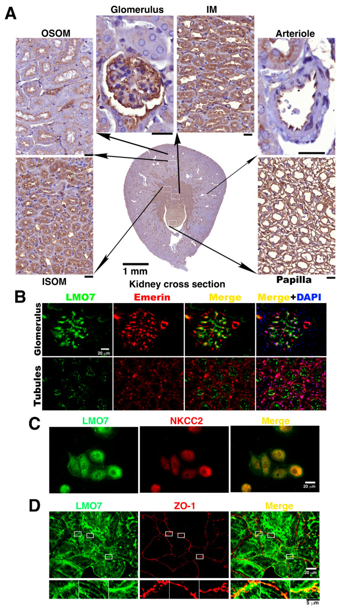
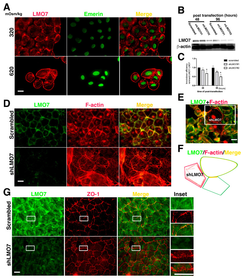
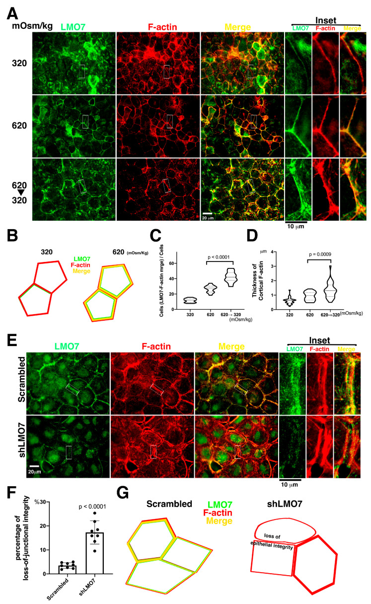
The kidney epithelial barrier has multifaceted functions in body fluids, electrolyte homeostasis, and urine production. The renal epithelial barrier (REB) frequently faces and challenges osmotic dynamics, which gives rise to osmotic pressure (a physical force). Osmotic pressure overloading can crack epithelial integrity and damage the REB. The endurance of REB to osmotic pressure forces remains obscure. LMO7 (LIM domain only 7) is a protein associated with the cell-cell junctional complex and cortical F-actin. Its upregulation was observed in cells cultured under hypertonic conditions. LMO7 is predominantly distributed in renal tubule epithelial cells. Hypertonic stimulation leads to LMO7 and F-actin assembly in the cortical stress fibers of renal epithelial cells. Hypertonic-isotonic alternation, as a pressure force pushing the plasma membrane inward/outward, was set as osmotic disturbance and was applied to test FAK signaling and LMO7 functioning in maintaining junctional integrity. LMO7 depletion in cells resulted in junctional integrity loss in the epithelial sheet-cultured hypertonic medium or hypertonic-isotonic alternation. Conversely, FAK inhibition by PF-573228 led to failure in robust cortical F-actin assembly and LMO7 association with cortical F-actin in epithelial cells responding to hypertonic stress. Epithelial integrity against osmotic stress and LMO7 and FAK signaling are involved in assembling robust cortical F-actin and maintaining junctional integrity. LMO7 elaborately manages FAK activation in renal epithelial cells, which was demonstrated excessive FAK activation present in LMO7 depleted NRK-52E cells and epithelial integrity loss when cells with LMO7 depletion were exposed to a hypertonic environment. Our data suggests that LMO7 regulates FAK activation and is responsible for maintaining REB under osmotic disturbance.

Keywords: FAK hypertonicity; LMO7; cortical stress fiber; epithelial barrier; epithelial integrity; osmotic stress; secretome.


Conflict of interest statement

All authors declare that they have no competing financial interest, such as consultancies, stock ownership or other equity interests or patent-licensing arrangements.

Figures

Figure 1
Figure 1
LMO7 expression in kidney. (A). LMO7 expression levels are relatively higher in renal cells in the inner stripe of the outer medulla (ISOM), inner medulla (IM), and papilla, and lower in renal cells in the outer stripe of the outer medulla (OSOM). In addition to renal tubules, LMO7 is also detected in the glomerular endothelial cells lining arterioles and podocytes. Bars, 20 µm. (B). To visualize the LMO7 subcellular localization in glomeruli and nephritic ducts, kidney specimens are stained with an antibody against LMO7 (green) and an antibody recognizing emerin (red), which is an inner nuclear protein and is characterized as a LMO7 binding partner. LMO7 is detected in the glomerulus and apical portion of the renal tubular cells. (C). In NKCC2 positive renal tubular epithelial cells, LMO7 is present in the periphery of cells and boundaries of two junctional cells. (D). In NRK-52E cells, LMO7 associates with zonula occludens-1 (ZO-1), a tight junction protein.
Figure 2
Figure 2
LMO7 associated with peripheral stress fibers in NRK-52E cells. (A). LMO7 distributions in NRK-52E epithelial sheets are visualized with antibody-recognized LMO7 (Red) and emerin (Green). In the NRK-52E epithelial sheet, LMO7 forms fiber-like structures and is distributed in the cell peripheries or transversely in the cytoplasm. When cells are exposed to 620 mOsm/kg for 24 h, they are longer in an epithelial sheet, and individual cells isolated from the epithelial sheet are discerned. LMO7 cells cultured in hypertonic medium for 24 h are present in the cell peripheries in a circular style. Bar = 20 µm. (B). LMO7 depletion by small hairpin RNA (shRNA) in NRK-52E cells is performed using transient shRNA transfection. Two distinct shRNAs targeting the LMO7 transcript are used to knock down LMO7 expression in NRK-52E cells. shLMO7#1 and shLMO7#2 demonstrate different LMO7 depletion efficiencies. By shRNA transfection, more than half of LMO7 reduces in NRK-52E cells with shLMO7#2, compared to LMO7 expression in NRK-52E cells with scrambled shRNA targeting luciferase. (C). Protein band densities are evaluated, and LMO7 protein band density from scrambled is defined as one unit to evaluate other LMO7 band densities. * p < 0.05 compared with Scrambled shRNA transfected cells. (D). As visualized by F-actin organization in NRK-52E and LMO7 depleted NRK-52E cells, peripheral stress fibers formed in NRK-52E cells are absent in LMO7 depleted cells. In NRK-52E cells, LMO7 associates with cortical stress fibers in epithelial sheet-like NRK-52E cells. (E). In NRK-52E cells with different LMO7 expression levels, robust peripheral F-actin cables assemble in cells with higher LMO7 expression and disordered F-actin fibers distributes in the cells with lower LMO7 expression levels. (F). A schematic representation of the three cells in the upper right corner of Figure F displays LMO7 merged with robust F-actin cables in cell peripheries and the left cell harbors disordered fine F-actin fibers in the cells harboring few LMO7. (G). LOM7 depleted NRK-52E cells and the cells without LOM7 depletion were cultured in 620 mOsm/kg medium for two hours. Zonula occludens-1 (ZO-1) (Red) and LMO7 (Green) in cells were visualized with antibodies, respectively labeled. By osmotic stress, the thicker ZO-1 formed in boundaries of two cells in NRK-52E cells without LMO7 depletion. In LMO7 depleted NRK-52E cells, ZO-1 are aberrantly distributed in boundaries of two cells.
Figure 3
Figure 3
Effect of LMO7 depletion on junctional integrity is susceptible to osmotic pressure challenge. (A). The epithelial sheets are cultured in isotonic, hypertonic, and 620/320 mOsm/kg alternating medium. F-actin structures (Red) and LMO7 (Green) distributions are visualized in the epithelial sheet. LMO7 partly associates with fine cortical stress fibers in epithelial sheets cultured in isotonic medium (320 mOsm/kg) for 2 h, and epithelial integrity is preserved. When epithelial sheets are exposed to a hypertonic milieu for 2 h, thickening cortical stress fibers are assembled, and LMO7 proteins associate with robust cortical stress fibers. When the epithelial sheets are confronted with 620/320 mOsm/kg alternated osmolarity (620 mOsm/kg for hour, and 320 mOsm/kg for another hour), cortical stress fibers and their parallel stress fibers assemble in cell peripheries in epithelial-sheet and LMO7 localizes with those stress-fibers. (B). Schematic representation demonstrates that thin cortical stress fibers present in the epithelial cell cortex under isotonic 320 mOsm/kg and LMO7 partly associate with cortical F-actin. Exposing to 620mOsm/kg, thicker cortical F-actin assemble in the renal epithelial peripheries, and LMO7 associated with cortical F-actin. (C). Hypertonic stimulation promotes LMO7 expression associated with cortical F-actin. The number of cells harboring LMO7/F-actin assembled cortical cytoskeletons is increased in cells grown in hypertonic medium and which undergo hypertonic-isotonic alternation. (D). Cortical F-actin fibers assemble in thicker cable when epithelial cells grow at 620 mOsm/kg and undergo 620-to-320 mOsm/kg alternation. (E). The epithelial sheets are exposed to osmotic challenge at 620/320 mOsm/kg alternated osmotic milieu and F-actin organization, and LMO7 expression is examined in renal epithelial sheet individual cells. After LMO7 depletion, epithelial integrity is obviously damaged and disordered cortical actin stress fiber organization is visible in the cell peripheries. Additionally, epithelial integrity loss is visible, and a gap between two adjacent cells and junctional connections loss with two parallel cortical stress fibers in cell peripheries are observed. (F). The cells that do not tightly adhere together in the visualized view are counted as cells with junctional integrity loss. Most cells in the renal epithelial sheet that undergo hypertonic-to-isotonic alternation are tightly adhered together, and few are not junctional to its adjacent cells in the NRK-52E epithelial sheet. When the LMO7 depleted NRK-52E epithelial sheet is challenged with hypertonic-isotonic alternation, osmotic disturbance causes severe junctional integrity loss in the LMO7 depleted NRK-52E epithelial sheet. (G). The schematic diagram displays LMO7 associated with cortical F-actin in cell cortices, and the cells are tightly attached to their adjacent cells. When LMO7 depletes in epithelial cells, they lose their junctional ability to adhere to their adjacent cells.
Figure 4
Figure 4
Osmotic induction leads to FAK phosphorylation and its localization at focal adhesion. (A). Focal adhesion kinase (FAK) phosphorylated on tyrosine 576/577 (p-FAK) is validated in NRK-52E cells exposed to various 620 mOsm/kg hypertonic medium durations. p-FAK reaches the highest level at 15 min after 620 mOsm/kg exposure. Subsequently, p-FAK levels abruptly decrease. One hour after hypertonic stimulation, p-FAK levels in NRK-52E cells are similar to those in cells without osmotic stimulation. After hypertonic stimulation for 4 h, p-FAK levels remain almost constant in NRK-52E cells. (B). The p-FAK levels in NRK-52E cells treated with 620 mOsm/kg hypertonic duration are plotted in a time-dependent curve. The p-FAK levels are highest in cells exposed to 620 mOsm/kg hypertonic medium for 15 min. (C). LMO7 expression in NRK-52E cells exposed to 620 mOsm/kg were evaluated by Western blot analysis. (D). The LMO7 protein band densities were digitalized and normalized to corresponding β-actin bands. Ratio of LMO7 to β-actin were plotted in a time-dependent curve. (E). p-FAK translocates to focal adhesions in NRK-52E cells cultured in 620 mOsm/kg hypertonic medium.
Figure 5
Figure 5
Fine peripheral stress fibers, less LMO7, and junctional integrity loss are present in FAK inhibitor-administrated cells in epithelial sheet. (A). The enzymatic function of focal adhesion kinase is blocked by PF-573228. The LMO7 (Green) in NRK-52E cells in epithelial sheet-like cells is fiber-like and is associated with peripheral F-actin (Red) stress fibers or transverses in the cytoplasm without osmotic induction and FAK inhibition. When exposed to hypertonic conditions, cortical F-actin stress fibers assemble in the periphery of cells in an epithelial sheet-like manner, and LMO7 localizes with the cortical F-actin stress fibers. When PF-573228 is present in culture in normal or hypertonic DMEM, the fine cortical F-actin stress fibers in the periphery of cells in the epithelia sheet and LMO7 also represent fine fiber-like structures. FAK inhibition attenuates robust cortical F-actin stress fiber assembly and reduces LMO7-associated cortical F-actin stress fibers in NRK-52E cells. (B). The cells are stained with antibodies against LMO7 (Green) and β-catenin (Red). Epithelial sheets settle in an osmotic disturbance altered by the medium osmolarity, which is 620 mOsm/kg hypertonic medium for an hour, followed by 320 mOsm/kg isotonic medium for another hour, which is designated as 620/320 mOsm/kg. Without PF-573228 presence, LMO7 forms a fiber-like structure parallel to the linear β-catenin at the boundary of two cells in the epithelial sheet, confronting osmotic disturbance. When the epithelial sheets are cultured in osmotic disturbance in the presence of PF-573228, parallel LMO7 fibers are absent, and aberrant β-catenin distribution is visible in the boundary between the two cells in the epithelial sheet. Inhibiting FAK enzymatic activity leads to junctional integrity loss in the NRK-52E epithelial sheet in osmotic disturbance. (C). FAK activation by osmotic disturbance leads to robust cortical F-actin assembly in cell peripheries and establishes compacted cell–cell junctions in the renal epithelial sheet. The inhibition of FAK enzymatic activity by PF-573228 results in fine cortical F-actin assembly in cells cultured in 320 or 620 mOsm/kg medium. (D). Number of cells with lost cell-cell junction and number of total cells in image were counted respectively. Number of cells with lost cell-cell junction divided by total cell number were calculated as percentage of loss-of-junctional integrity. Approximate 2.4% cells do not bear completed junctions in NRK-52E epithelial sheet exposed to 620 mOsm/kg without presence of PF-573228. When FAK inhibitor was added to 620 mOsm/kg hypertonic medium, 12.8% of total cells lost junctional integrity.
Figure 6
Figure 6
Junctional integrity loss and excessive FAK phosphorylation in LMO7 depleted NRK-52E depleted epithelial sheet, which underwent alternated osmotic challenge. (A). The epithelial sheet undergoes hypertonic-isotonic alternation for an hour. Cells in the epithelial sheets are stained with antibodies against p-FAK (Green) and Alexa-568 conjugated phalloidin (Red). In the NRK-52E epithelial sheet without LMO7 depletion, p-FAK is present at focal adhesions (FA), and cortical actin assembles around the cell cortex. The hypertonic-isotonic alternation does not compromise epithelial integrity, and no junctional integrity loss is observed. Conversely, relatively more p-PTK-based FAs assemble and disordered F-actin stress fibers assemble in LMO7 depleted NRK-52E epithelial sheets under hypertonic-isotonic alternation. (B). p-FAK levels are higher in LMO7 depleted NRK-52E cells cultured in the medium with 620 mOsm/kg. (C). Band densities are digitalized and the ratio of p-FAK to β-actin is calculated. This indicates that p-FAK levels are elevated in LMO7 depleted cells with or without 620 mOsm/kg hypertonic stimulation. (D). NRK-52E cells with/ without LMO7 depletion were stained with antibodies, respectively recognized LMO7 (Green) and paxillin phosphorylation on tyrosine 31 designated as p-Paxillin (Red). Without LMO7 depletion, the p-Paxillin based focal adhesions (FAs) are randomly distributed in cells when cells were expose in 620 mOsm/kg. In the LMO7 depleted cells, the 620 mOsm/kg osmotic stimulation resulted in more p-Paxillin present in FAs, and specifically the p-Paxillin based FAs predominately distributed boundary of two cells.
Figure 7
Figure 7
FAK inhibition attenuates osmotic tolerance of NRK-52E cells and osmotic disturbance causes cell death. (A). Cells are cultured in 320 mOsm/kg isotonic and hypertonic media at 420, 520, and 620 mOsm/kg for 24 h. In isotonic or 420 mOsm/kg medium, few dead cells are detected using an apoptotic assay with flow cytometry. It appears that 620 mOsm/kg hypertonic stress damages NRK-52E cells and causes cell death. (B). FAK signaling regulates cell survival under hypertonic stress. FAK inhibition leads to cell death by hypertonic shock. (C). Schematic representation depicts that LMO7 facilitates cortical F-actin stress fiber assembly under hypertonic shock to sustain epithelial integrity. Under hypertonic stress, osmotic pressure pushes the plasma membrane inward and activates FAK. The FAK catalytic activity regulates cortical F-actin reassembly around cell peripheries in the epithelial sheet. LMO7 is an important element incorporated into cortical stress fibers and ordered robust microfilament organization around the cell peripheries. Subsequently, this robust F-actin/LMO7 structure attenuates FAK activation. When LMO7 is absent in epithelial cells, disordered cortical F-actin structure, instead of robust cortical F-actin stress fibers, appear in cell peripheries. The epithelial sheet is fragile when epithelial cells bear disordered cortical F-actin fibers. This disordered F-actin organization in the epithelial cells fails to resist osmotic pressure. As consequence, osmotic pressure force continues to activate FAK, and excessive active FAK promotes pathological epithelial-mesenchymal transition (EMT) and cell migration. Epithelial integrity loss appears in LMO7 depleted epithelial sheet.

Similar articles

Cited by

References

    1. Dantzler W.H., Layton A.T., Layton H.E., Pannabecker T.L. Urine-concentrating mechanism in the inner medulla: Function of the thin limbs of the loops of Henle. Clin. J. Am. Soc. Nephrol. 2014;9:1781–1789. doi: 10.2215/CJN.08750812. - DOI - PMC - PubMed
    1. Hara M., Minami Y., Ohashi M., Tsuchiya Y., Kusaba T., Tamagaki K., Koike N., Umemura Y., Inokawa H., Yagita K. Robust circadian clock oscillation and osmotic rhythms in inner medulla reflecting cortico-medullary osmotic gradient rhythm in rodent kidney. Sci. Rep. 2017;7:7306. doi: 10.1038/s41598-017-07767-8. - DOI - PMC - PubMed
    1. Di Ciano C., Nie Z., Szaszi K., Lewis A., Uruno T., Zhan X., Rotstein O.D., Mak A., Kapus A. Osmotic stress-induced remodeling of the cortical cytoskeleton. Am. J. Physiol. Cell Physiol. 2002;283:C850–C865. doi: 10.1152/ajpcell.00018.2002. - DOI - PubMed
    1. Roffay C., Molinard G., Kim K., Urbanska M., Andrade V., Barbarasa V., Nowak P., Mercier V., Garcia-Calvo J., Matile S., et al. Passive coupling of membrane tension and cell volume during active response of cells to osmosis. Proc. Natl. Acad. Sci. USA. 2021;118:e2103228118. doi: 10.1073/pnas.2103228118. - DOI - PMC - PubMed
    1. Romero S., Le Clainche C., Gautreau A.M. Actin polymerization downstream of integrins: Signaling pathways and mechanotransduction. Biochem. J. 2020;477:1–21. doi: 10.1042/BCJ20170719. - DOI - PubMed

LinkOut - more resources