. 2022 Nov 29;11(23):3834.
doi: 10.3390/cells11233834.

Delivery of Active AKT1 to Human Cells

Affiliations

Delivery of Active AKT1 to Human Cells

Tarana Siddika et al. Cells. .

Abstract

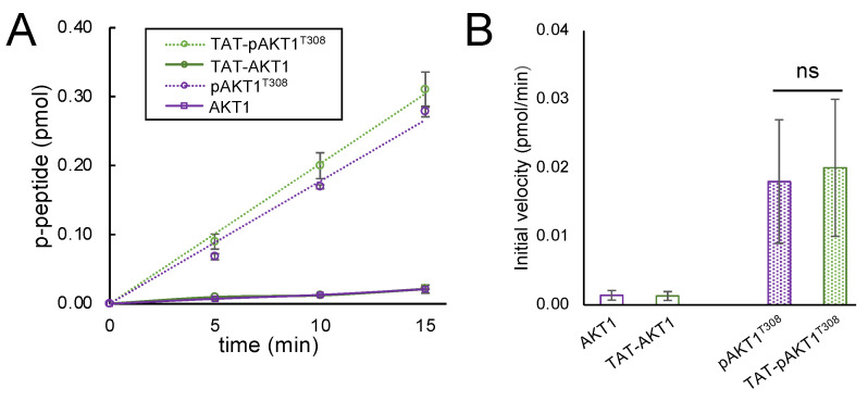
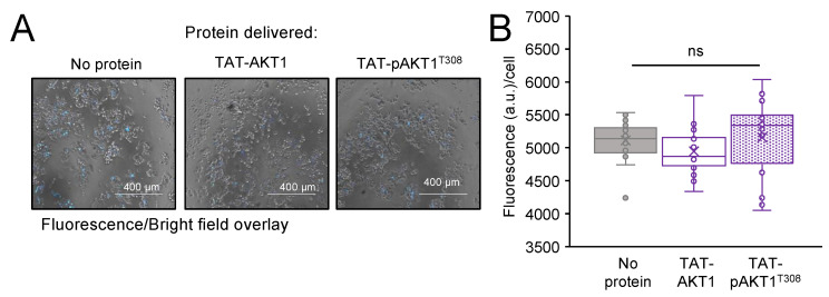
Protein kinase B (AKT1) is a serine/threonine kinase and central transducer of cell survival pathways. Typical approaches to study AKT1 biology in cells rely on growth factor or insulin stimulation that activates AKT1 via phosphorylation at two key regulatory sites (Thr308, Ser473), yet cell stimulation also activates many other kinases. To produce cells with specific AKT1 activity, we developed a novel system to deliver active AKT1 to human cells. We recently established a method to produce AKT1 phospho-variants from Escherichia coli with programmed phosphorylation. Here, we fused AKT1 with an N-terminal cell penetrating peptide tag derived from the human immunodeficiency virus trans-activator of transcription (TAT) protein. The TAT-tag did not alter AKT1 kinase activity and was necessary and sufficient to rapidly deliver AKT1 protein variants that persisted in human cells for 24 h without the need to use transfection reagents. TAT-pAKT1T308 induced selective phosphorylation of the known AKT1 substrate GSK-3α, but not GSK-3β, and downstream stimulation of the AKT1 pathway as evidenced by phosphorylation of ribosomal protein S6 at Ser240/244. The data demonstrate efficient delivery of AKT1 with programmed phosphorylation to human cells, thus establishing a cell-based model system to investigate signaling that is dependent on AKT1 activity.

Keywords: cell penetrating peptide; cellular signaling; kinase; phosphoinositide-dependent kinase (PDK1); protein kinase B (AKT1); recombinant protein; trans-activator of transcription (TAT).


Conflict of interest statement

The authors declare no conflict of interest.

Figures

Figure 1
Figure 1
AKT1 activation pathway and delivery of site-specifically phosphorylated AKT1 into human cells. (A) Protein kinase B (AKT1) is normally activated in response to growth factors that bind to a receptor tyrosine kinase (RTK) in the membrane, activating phosphoinositide 3-kinase (PI3K). PI3K subsequently phosphorylates phosphatidylinositol-4,5-bisphosphate (PIP2) to phosphatidylinositol-3,4,5-trisphosphate (PIP3). When the pleckstrin homology (PH) domain of AKT1 binds to the membrane phospholipid PIP3, the auto-inhibitory effect of the PH domain is released from AKT1, exposing the activation sites of AKT1 for phosphorylation by the up-stream kinases mTOR complex 2 (mTORC2) at Ser473 and phosphoinositide-dependent kinase 1 (PDK1) phosphorylates Thr308. The activation of AKT1 initiates a phosphorylation cascade, leading to the activation of many downstream pathways controlling cell growth and protein synthesis as well as inhibition of apoptosis. (B) Trans-Activator of Transcription (TAT) is a cell penetrating peptide facilitating protein delivery to mammalian cells. TAT-tagged AKT1 was expressed and purified from E. coli cells. PDK1 was co-expressed in E. coli to facilitate phosphorylation of AKT1 at Thr308 during protein production, yielding active, phosphorylated TAT-tagged AKT1. TAT-tagged AKT1 variants were then delivered to human cells.
Figure 2
Figure 2
Enzymatic activity of AKT1 is dependent on phosphorylation at Thr308 and was not affected by fusion of AKT1 to the cell penetrating TAT-tag. (A) Time courses and (B) initial velocity of three independent in vitro kinase activity assays. AKT1 variants were incubated with a GSK-3β substrate peptide and [γ-32P]-ATP and spotted on filter paper. Unreacted [γ-32P]-ATP was washed away and reaction products visualized by phosphorimaging (Figure S3) and quantified. AKT1 (purple) and TAT-AKT1 (green) did not show significant activity above background, whereas pAKT1T308 (purple dotted) and TAT-pAKT1T308 (green dotted) were enzymatically active. The TAT-tag did not significantly alter pAKT1T308 activity. Significant differences were calculated by two-tailed t-test (ns—not significant).
Figure 3
Figure 3
Delivery of TAT-AKT1 protein variants to HEK 293T cells. HEK 293T cells were incubated with AKT1, pAKT1T308, TAT-AKT1, or TAT-pAKT1T308 for 1 h in three biological replicates. Cell extracts were separated by SDS-PAGE and immunoblotted with AKT1, pAKTT308, and GAPDH specific antibodies. Cells incubated with AKT1 or pAKT1T308 showed a single band in all blots corresponding to endogenous AKT1. Two bands, corresponding to endogenous AKT1 and TAT-AKT1 separated by size, were apparent in cells transfected with TAT-tagged AKT1 variants, demonstrating effective delivery of both TAT-tagged AKT1 variants into cells. TAT-AKT1 delivery did not lead to phosphorylation of TAT-AKT1 in cells, as no phosphorylated TAT-AKT1 was detected by the pThr308 specific antibody after 1 h. The delivered TAT-pAKT1T308 was readily detected by phospho-specific antibody for pAKTT308.
Figure 4
Figure 4
TAT-pAKT1T308 protein delivery stimulates AKT1 signaling. HEK 293T cells were incubated with AKT1, pAKT1T308, TAT-AKT1, or TAT-pAKT1T308 for 1 h in three biological replicates. (A) Cell extracts were separated by SDS-PAGE and immunoblotted with GSK-3, pGSK-3, and GADPH specific antibodies. GSK-3 homologs GSK-3α and GSK-3β are direct substrates of AKT1. (B) Quantification of western blots showed no change in GSK-3α or GSK-3β protein abundance. Changes in phosphorylation of GSK-3α or GSK-3β were quantified and normalized to (C) GAPDH or (D) GSK-3α and GSK-3β, respectively, showing that TAT-pAKT1T308 specifically stimulates phosphorylation of GSK-3α, but not GSK-3β. Significant differences were calculated by two-tailed t-test and are indicated by asterisks (** p < 0.01).
Figure 5
Figure 5
TAT-pAKT1T308 protein delivery stimulates downstream AKT1 signaling to ribosomal protein S6. HEK 293T cells were incubated with AKT1, pAKT1T308, TAT-AKT1, and TAT-pAKT1T308 for 1 h in three biological replicates. (A) Western blots and (B,C) quantification of blots of cell extracts separated by SDS-PAGE and immunoblotted with ribosomal protein S6, pS6, and GADPH specific antibodies. S6 phosphorylation was significantly increased after incubation with TAT-pAKT1T308, but not following incubation of cells with AKT1 or pAKT1 lacking the TAT tag or with unphosphorylated TAT-AKT protein variants. Significant differences were calculated by two-tailed t-test and are indicated by asterisks (* p < 0.05).
Figure 6
Figure 6
AKT1 over-expression and growth factor stimulation activates AKT1 signaling in HEK 293T cells. HEK 293T cells were transfected with an mCherry-AKT1 over-expression plasmid. At 24 h after transfection, cells were stimulated with EGF for 10 min. (A) Cell extracts were separated by SDS-PAGE and immunoblotted with AKT1, GSK-3, pGSK-3, and GAPDH specific antibodies in three biological replicates. (B) Quantification of western blots showed no change in GSK-3α or GSK-3β protein abundance. (C) GSK-3α and GSK-3β phosphorylation normalized to GAPDH was significantly increased in cells over-expressing AKT1 after EGF stimulation, but not significantly changed in EGF stimulated cells alone or unstimulated cells over-expressing AKT1. (D) GSK-3α and (E) GSK-3β phosphorylation was also normalized to GSK-3α and GSK-3β levels, showing increased phosphorylation of both GSK-3 isoforms in response to AKT1 over-expression, which was further increased upon EGF stimulation. Error bars represent the standard deviation of the mean. p-values were calculated by two-tailed t-test and are indicated by asterisks (* p < 0.05, ** p < 0.01).
Figure 7
Figure 7
Delivery of TAT-mCherry-AKT1 variants compared to plasmid-based transfection efficiency of mCherry-AKT1. (A) Overlay of brightfield and fluorescent (excitation 531 nm, emission 593 nm) images (above) and zoomed-in images (below) of cells 24 h after incubation with TAT-mCherry-AKT1, TAT-mCherry-pAKT1T308, or lipofectamine mediated transfection with an mCherry-AKT1 expressing plasmid. (B) Quantification of delivery or transfection efficiency from three biological replicates was calculated as the ratio of transfected cells/total cells and by determining fluorescence of the mCherry-AKT1 fusion proteins in cells. Protein delivery and plasmid transfection efficiency was not significantly different. The data are based on three biological replicates, error bars show ±1 standard deviation, and significance was calculated by one-way ANOVA (ns—not significant).
Figure 8
Figure 8
TAT-AKT1 protein delivery is non-toxic to HEK 293T cells. Cytotoxicity was measured after a 24-h incubation with no protein (buffer only), TAT-AKT1, or TAT-pAKT1T308 by staining dead cells with Sytox blue in three biological replicates. (A) Images of cells with Sytox blue staining of dead cells. (B) Quantification of dead cells shows no significant impact of TAT-AKT1 and TAT-pAKT1T308 protein delivery compared to cells treated with buffer only (no protein). Significance was calculated by one-way ANOVA (ns—not significant).

Similar articles

Cited by

References

    1. Vogiatzi P., Giordano A. Following the tracks of AKT1 gene. Cancer Biol. Ther. 2007;6:1521–1524. doi: 10.4161/cbt.6.10.4834. - DOI - PubMed
    1. Chen W.S., Xu P.-Z., Gottlob K., Chen M.-L., Sokol K., Shiyanova T., Roninson I., Weng W., Suzuki R., Tobe K. Growth retardation and increased apoptosis in mice with homozygous disruption of the Akt1 gene. Genes Dev. 2001;15:2203–2208. doi: 10.1101/gad.913901. - DOI - PMC - PubMed
    1. Karege F., Perroud N., Schürhoff F., Meary A., Marillier G., Burkhardt S., Ballmann E., Fernandez R., Jamain S., Leboyer M. Association of AKT1 gene variants and protein expression in both schizophrenia and bipolar disorder. Genes Brain Behav. 2010;9:503–511. - PubMed
    1. Santi S.A., Lee H. The Akt isoforms are present at distinct subcellular locations. Am. J. Physiol. -Cell Physiol. 2010;298:C580–C591. doi: 10.1152/ajpcell.00375.2009. - DOI - PubMed
    1. Staal S.P. Molecular cloning of the akt oncogene and its human homologues AKT1 and AKT2: Amplification of AKT1 in a primary human gastric adenocarcinoma. Proc. Natl. Acad. Sci. USA. 1987;84:5034–5037. doi: 10.1073/pnas.84.14.5034. - DOI - PMC - PubMed

MeSH terms